Biology 2382B Lecture Notes - Lecture 24: Programmed Cell Death, Differential Interference Contrast Microscopy, Mitochondrion

2 Apr 2016
School
Department
Course
Professor
Document Summary
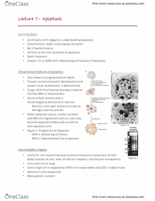
Apoptosis (also known as programmed cell death) is a key process that ensures normal embryonic development and proper issue homeostasis in adult animals. Programmed cell death is an acive cellular process which requires energy as opposed to necrosis (the passive form of cell death) where cells simply swell up and die. One of the key features of apoptosis is chromain condensaion as the nucleus starts to morphological changes. Apoptoic cells tend to shrink somewhat as well, and blebs (irregular bulges in plasma membrane) start to become visible. As this progresses, the blebbing coninues and the nucleus actually breaks down in smaller fragments known as apoptoic bodies, which are recognized and engulfed whole by phagocyic cells in order to minimize cellular debris produced. Apoptosis also involves the cleavage of dna by dnaases (icad) that cut in between nucleosomes (periodicity of about 200 base- pairs), yielding 200 base-pair fragments.