Health Sciences 3300A/B Lecture 14: Lecture 14 - Abodminal Cavity & Stomach

19 Feb 2019
School
Department
Course
Professor

Lecture 14 - Abodminal Cavity & Stomach
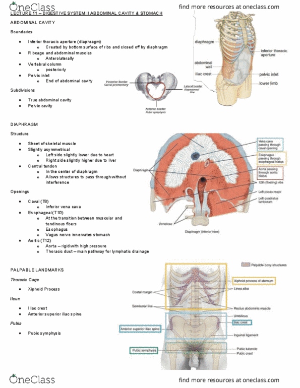
Abdominal Cavity
- Inferior thoracic aperture is created by the bottom surface of the ribs - closed off by the
muscular diaphragm
- The rib cage and muscles (anterior-laterally), vertebral column (posteriorly) and the pelvic inlet
(end of the abdominal cavity)
- The pelvic cavity has its own section within the abdominal space
- The ilopectineal line creates a smooth edge - travels around the sacral promontory and
finishes off at the pubic symphysis
- Everything below this line is the true pelvis, everything above the line but within the fossa is
the false pelvis
Diaphragm
- Can be activated voluntarily, but does have automatic innervation as well
- The left side is slightly lower and the right side is slightly higher due to the liver that pushes up
the diaphragm and the heart pushes down on the diaphragm
- As you move towards the center, it becomes tendonous
→ Allows structure to pass through without interference from the diaphragmatic
contractions
- Esophageal origin is at the transition b/w the muscular and tendonous fibers
→ Vagus nerve passes the diaphragm and innervates the stomach
- The aorta is rigid w/ high pressure, so it has no problem staying open
- Thoracic ducts are the main pathway for lymphatic drainage
Abdominal Quadrants
- The largest cavity in the body - must be divided into four quadrants
- Transumbilical plane (horizontal - L4) and median plane (travels vertically down the midline)
- The liver is on the right-hand side, but the left lobe goes a bit past the median plane
Abdominal Regions
- Medically and surgically, we use regions which give us more precise locations
- Regions exist in 9 pieces & are divided by two vertical lines and horizontal lines
- Midclavicular planes cross over the 9th costal cartilage and travel in between the pubic
symphysis and the ASIS
- Epigastric region is right above the stomach
Peritoneum
- Similar to the lining in the respiratory system
- Parietal peritoneum lies the walls of the cavity
- Visceral peritoneum covers most of the organs in the abdominal cavity
- If an abdominal cavity is moved around, you only see the organs within the peritoneal cavity -
everything behind the membrane is hidden
Document Summary
Inferior thoracic aperture is created by the bottom surface of the ribs - closed off by the muscular diaphragm. The rib cage and muscles (anterior-laterally), vertebral column (posteriorly) and the pelvic inlet (end of the abdominal cavity) The pelvic cavity has its own section within the abdominal space. The ilopectineal line creates a smooth edge - travels around the sacral promontory and finishes off at the pubic symphysis. Everything below this line is the true pelvis, everything above the line but within the fossa is the false pelvis. Can be activated voluntarily, but does have automatic innervation as well. The left side is slightly lower and the right side is slightly higher due to the liver that pushes up the diaphragm and the heart pushes down on the diaphragm. As you move towards the center, it becomes tendonous. Allows structure to pass through without interference from the diaphragmatic contractions.