Kinesiology 1080A/B Lecture 18: Kin lecture 18 - March 1

8 Mar 2017
School
Department
Course
Professor
Document Summary
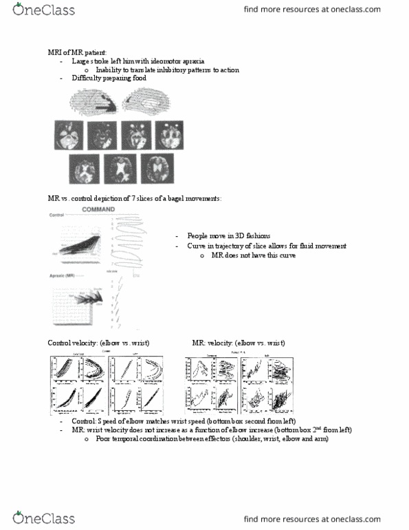
Poor temporal coordination between his effectors: measuring cns activity, electromyography. Provides information regarding the temporal and intensity characteristics of the movement: reciprocal innervation agonist muscle is active; antagonist muscle is inactive, tonic activation, what emg looks like if you electrically stimulate the muscle. You get synchronist or overlapping of activity of extrafusal muscle fibers they are all firing at the exact same time. From an electrical or magnetic input: phasic activation, what emg looks like for voluntary contraction, with a voluntary contraction, you get a synchronist activity the (cid:373)ove(cid:373)e(cid:374)ts do(cid:374)(cid:859)t overlap entirely in time (they can be close) Signals doing get to the muscle fibers at the same time; asynchronally activating extrafusal muscle fibers. Provide measures of when and what brain regions activate during movement. Positron emission tomography (pet) and functional magnetic resonance imaging (fmri) Pet metabolic activity in the brain; glucose uptake: terrible special and temporal resolution; measure every 10 seconds.