Kinesiology 1080A/B Lecture Notes - Lecture 14: Motor Neuron, Reciprocal Inhibition, Corticobulbar Tract

13 Mar 2019
School
Department
Course
Professor
Document Summary
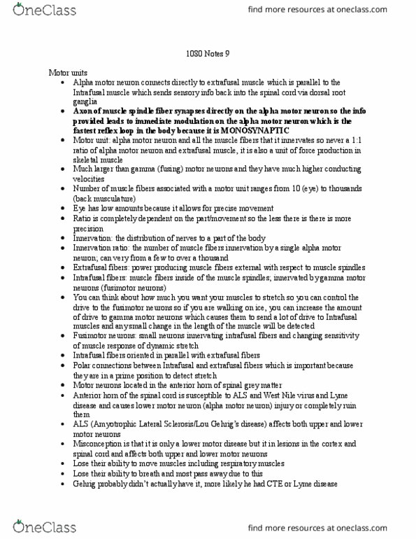
Ventral root and ventral root ganglion: motor commands leave here. Green circles represents the cell body of an alpha motor neuron. That alpha neuron is going to innervate the extrafusal muscle fiber. Causing a contraction and the body to move. Golgi tendon organs gto: monitor for force in an extrafusal muscle fiber. *monosynaptic stretch reflex depicted by an alpha motor neuron is going to innervate the extrafusal muscle fiber. Interneuron (red, its inhibitory) will intern connect to an alpha motor neuron telling it to stop firing so stop tension. Interneurons: exist in one level of the spinal cord, connect neurons together. Propriospinal neurons: different from interneurons, connect neurons in the spinal cord, but have to travel multiple lengths within the spinal cord (so longer) Pyramids: in medulla where descending motor fibers cross over to the other side, this is pyramidal decussation (crossing over point) approx. 70-80% of motor fibers cross over at this point, approx.