Physiology 3120 Lecture Notes - Sliding Filament Theory, Intercalated Disc, Sinoatrial Node

26 Nov 2011
School
Department
Course
Professor
Document Summary
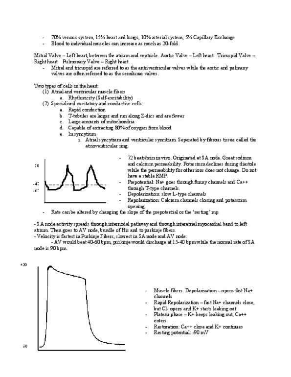
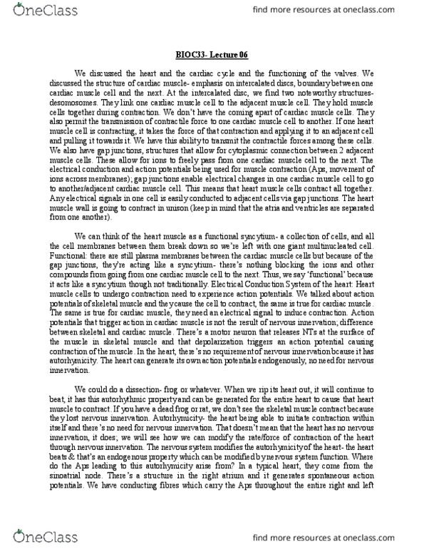
Function of myocardial cells: looking at microscopic level (cellular, molecular, etc. , heart beat = most important life-sustaining event; heart is first organ formed during embryogenesis, three types of muscle fibres, atrial*, ventricular*, excitatory & conductive. In skeletal muscle, muscle fibres function independently of each other; cardiac muscle fibres are interconnected & form a system (i. e. syncytium) Where two fibres meet, membrane folds in region called intercalated disc. Two functional syncytium in heart: atrial syncytium, contract before the ventricles because of the impulse generated in the sa node (must conduct down into ventricles, ventricular syncytium. **have contractile properties like skeletal muscle (i. e. sliding filament theory; however, there are major differences [i. e. troponin])** Impulse generated at sinus node (located just under vena cava in right atrium) Impulse generation in this area occurs faster than elsewhere in the heart. Special properties (all of which work to depolarize the sa node cells) K permeability declines during diastole (more positive charges inside cell)