Physiology 2130 Lecture Notes - Lecture 10: Globin, Arteriole, Reversible Reaction

17 Jan 2017
School
Department
Course
Professor

Physiology 10 (10.1-10.82)
Respiratory System
Introduction
Functions:
o Transport of oxygen from air into blood
o Removal of carbon dioxide from blood into air
o Control of blood acidity (pH)
o Temperature regulation
o Forming a line of defense to airborne particles
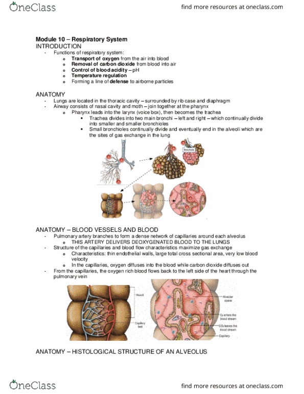
Anatomy
Located in the thoracic cavity
Surrounded by the rib cage and diaphragm,
Consists of nasal cavity and mouth – which join at the pharynx
Pharynx leads into the larynx (aka voice box) which then becomes the trachea
Trachea divides into two main bronchi (left and right)
Bronchi divide into smaller and smaller bronchioles
Bronchioles keep dividing into alveoli (site of gas exchange)
Pic on right
o 1) Larynx
o 2) trachea
o 3) right and left bronchi
o 4) right lung
o 5) left lung
pic on right
o 1) right main bronchus
o 2) bronchioles
o 3) alveoli
o 4) alveoli
o 5) bronchiole
o 6) smooth muscle
o 7) alveolar space
o 8) capillary
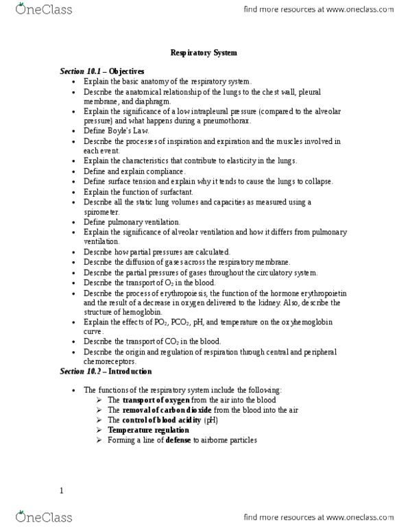
Anatomy – Blood Vessels
the pulmonary artery (which delivers deoxygenated blood to the lungs from the heart)
branches and forms a dense network of capillaries around each alveolus
capillaries have thin endothelial walls, large total cross-sectional area, and a very love blood
velocity
these characteristics help maximize gas exchange
SO in capillaries, oxygen diffuses into blood while carbon dioxide diffuses out
Now the oxygen rich blood flows back to left side of heart through pulmonary vein
Anatomy – Histological Structure of an Alveolus
There are about 300 million alveoli in healthy lung
Diameter: 0.3mm
Walls of alveoli are one cell thick and are composed of alveolar epithelial cells (type 1
cells)
Type 2 cells secrete a fluid called surfactant that lines the alveoli
Lots of capillaries surround alveoli
find more resources at oneclass.com
find more resources at oneclass.com

Region bw alveolar space and capillary lumen is the respiratory membrane
o Respiratory membrane is where gas exchange happens
o Can have thickness as narrow as 0.3 microns
Cells of immune system (called macrophages and lymphocytes) protect the body from
airborne particles that make their way into the alveoli
Fibers of elastin and collagen are present in the walls of the alveoli, around blood vessel and
bronchi
Pressures of the Lungs – Intrapleural Pressure
there are two thin pleural membranes
o one lines and sticks to the ribs (the parietal pleura)
o the other surrounds and sticks to the lungs (the visceral pleura)
these two layers of membrane form the intrapleural space which contains a small
amount of pleural fluid (about 10-15 ml)
the pleural fluid reduces friction bw the two pleural membranes during breathing
due to their nature and attached muscle, the ribs tend to spring outwards, while the lungs,
due to the presence of elastin, tend to recoil and collapse
Pressure of the Lungs – Alveolar and Atmospheric Pressure
the pressure inside the lungs is called the alveolar pressure (aka
intrapulmonary pressure)
the pressure in the intrapleural space is called the intrapleural pressure
the atmospheric pressure, outside the body, is 760 mmHg at sea level
bw breaths, the alveolar and atmospheric pressure are the same at 760mmHg (0 difference),
while the intrapleural space is about 756 mmHg (difference of 4)
the chest wall and lungs moving in opposite directions cause this lower intrapleural pressure
find more resources at oneclass.com
find more resources at oneclass.com

Pressure of the Lungs – Transpulmonary Pressure
transpulmonary pressure is the difference bw the alveolar and intrapleural pressure
the transpulmonary pressure is important bc this difference in pressure across the alveoli
and intrapleural space holds the lungs open
in a healthy set of lungs, the transpulmonary pressure is positive (outwards) and keeps the
lungs and alveoli open
Pressure of the Lungs - Pneumothorax
if alveolar pressure = intrapleural pressure, trans pulmonary pressure is 0mmHg
when its 0, there would be no pressure holding the lungs open and they would collapse,
producing a pneumothorax
this occurs when the intrapleural space is punctured, causing the alveolar pressure and
intrapleural pressure to become equal (both are 760 mmHg)
generally, only one lung collapses, bc the intrapleural space of each lung is isolated from the
other
Ventilation – Boyle’s Law
boyles law states that when the volume of a container decreases, the pressure inside
increases and vice versa
therefore, pressure is inversely proportional to volume
Ventilation – Inspiration and Expiration
moving air into the lungs requires a pressure gradient
this is called the air pressure gradient
to move air into the lungs, it requires high pressure outside and low pressure inside the
alveoli
to move air out requires high alveolar pressure and low atmospheric pressure
since you cant increase outside atmospheric pressure, the alveolar pressure must change
Ventilation – Mechanisms of Inspiration
to decrease the alveolar (intrapulmonary) pressure, the lung volume must increase
(according to boyles law)
to increase the volume, the diaphragm contracts, moving downwards, and the external
intercostal muscles of the rib contract, lifting the rib cage up and out
find more resources at oneclass.com
find more resources at oneclass.com
Document Summary
Functions: transport of oxygen from air into blood, removal of carbon dioxide from blood into air, control of blood acidity (ph, temperature regulation, forming a line of defense to airborne particles. Surrounded by the rib cage and diaphragm, Consists of nasal cavity and mouth which join at the pharynx. Pharynx leads into the larynx (aka voice box) which then becomes the trachea. Trachea divides into two main bronchi (left and right) Bronchi divide into smaller and smaller bronchioles. Bronchioles keep dividing into alveoli (site of gas exchange) Pic on right: 1) larynx, 2) trachea, 3) right and left bronchi, 4) right lung, 5) left lung. Pic on right: 1) right main bronchus, 2) bronchioles, 3) alveoli, 4) alveoli, 5) bronchiole, 6) smooth muscle, 7) alveolar space, 8) capillary. Anatomy blood vessels the pulmonary artery (which delivers deoxygenated blood to the lungs from the heart) branches and forms a dense network of capillaries around each alveolus.