Physiology 2130 Lecture Notes - Lecture 5: Podcast, Phosphate, Sliding Filament Theory

29 May 2018
School
Department
Course
Professor

Module 5 – Muscles
Intro:
• Muscles are biological machines that utilize chemical energy from the breakdown and metabolism of food to perform
useful work
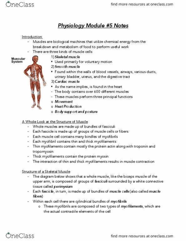
• There are 3 kinds of muscle cells
o Skeletal – used for voluntary muscle motion
▪ This module only covers skeletal muscles
o Smooth – found within the wall of blood vessels, airways, various ducts, urinary bladder, uterus and the
digestive tract
o Cardiac – found in the heart
• The body contains over 600 different muscles
• Muscles perform 3 principal functions:
1. Movement
2. Heat production
3. Body support and posture
Structure of a Skeletal Muscle:
• Descending in size
o Whole muscles are made up of bundles of fasciculi
▪ Which are surrounded by white connective tissue called perimysium
o Each fascicule is made up of groups of muscle cells or fibres
o Each muscle cell/fibre contains many bundles of myofibrils
o Each myofibril contains thin and thick myofilaments
o Thin myofilaments contain mostly the protein actin along with troponin and tropomyosin
o Thick filaments contain the protein myosin
• The interaction of thin and thick filaments results in muscle contraction
• The basic functional unit of a muscle is a sarcomere
A little ore i depth…
• Muscle cells/fibres are one of the few cells in the body with more than one nucleus
• They are surrounded by sarcolemma – the muscle cell membrane
o The AP is transmitted over this
• The sarcolemma has small tube-like projections called transverse tubules (or T tubules) that extend down the cell
o These T tubules conduct the AP deep into the cell where the contractile proteins are located
• With the muscle cell are long cylindrical myofibrils that contain the contractile proteins of the muscle
o The thin and thick filaments
• The myofibrils are surrounded by the sarcoplasmic reticulum (SR): a mesh like network of tubes containing calcium
ions (Ca+)
o Which are essential for contraction
• At either end of and continuous with the SR are the terminal cisternae: a membranous enlargement of the SR which is
close to the T tubule (where the AP travels)
Thin Myofilament:
• The thin myofilaments are composed predominantly of the globular protein actin
• Each actin molecule contains a special bind site for the other contractile protein myosin
• Many actin molecules are strung together like beads on a string to form the backbone
of the thin myofilaments
• Long strands of tropomyosin are found on the thin filaments
o These proteins cover the binding sites for myosin when the muscle is at rest
• Troponin is made up of three subunits
o Troponin A – binds to actin
o Troponin T – binds to tropomyosin
o Troponin C – binds to Ca+
find more resources at oneclass.com
find more resources at oneclass.com

Actin/Myosin Relationship:
• Groups of thin (actin) myofilaments and groups of thick (myosin) myofilaments are arranged in a repeating pattern
along the length of the myofibril from one end of the muscle to the other
• Each group of thin filaments extends outwards in opposite direction from the central Z disk or Z line
o Thin filaments are anchored to the Z line
• Each group of thick filaments extend outwards from a central M line, where they are anchored
• Each myofilament is parallel to the length of the myofibril and the muscle cell
• The region from one Z disk to another is called a sarcomere
o This is the smallest functional contractile unit of the muscle cell
• Under a microscope the repeating pattern of thin and thick filaments gives the muscle cell its banded/striated
appearance
o Regions of thick filaments are dark and called A bands
o Regions of thin filaments are lighter and called I bands
Muscle Contraction – The Sliding Filament Theory:
• The interaction between actin and myosin leads to muscle contraction
• Process
o When the head of the myosin molecule attaches to the binding site on actin and forms a cross bridge, the
myosin undergoes a change in shape
o This change in shape causes the myosin head to swing, producing the power stroke
o This power stroke propels or slides the actin filament past the myosin
• Note: the thick and thin filament do not change in size
Excitation-Contraction Coupling and Muscle Contraction:
• Excitation-Contraction Coupling: the process by which an AP in the cell
membrane excites the muscle cell to produce a muscle contraction
• The AP that was generated at the NMJ will spread out over the sarcolemma
and down the T-tubules into the core of the muscle cell
• The AP travels very close to the sarcoplasmic reticulum (SR) and will open Ca+
channels, causing the release of Ca+ from the terminal cisternae of the SR
• The Ca+ will bind to troponin C on the thin myofilaments, causing
tropomyosin to uncover the myosin binding sites found on action
• Myosin will now be able to attach to the actin and a power stroke will occur
Relaxation of Muscles:
• Once AP’s stop, Ca+ will no longer diffuse out of the sarcoplasmic reticulum (SR)
• Process:
o Special calcium pumps rapidly pump Ca+ back into the SR, up its concentration gradient – requires ATP
o Without Ca+ in the cytoplasm of the muscle cell the tropomyosin will cover the myosin binding sites again,
making myosin unable to bind and contraction unable to occur
o The muscle will then relax
• Important to note: active transport can be saturated – meaning that they can only work so quickly
o The removal of Ca+ is affected by this, meaning that a muscle may not be able to relax right away
Actin-Myosin and ATP Cycle:
• ATP is split to ADP and inorganic phosphate (Pi) which releases energy to
myosin and prepares the myosin head for activity
• Formation of the cross bridge occurs when Ca+, which have been released
from the SR by an AP, binds to tropomyosin C
• This rolls the tropomyosin off the myosin binding site on actin
• The power stroke occurs when the myosin head bends and slides the thin
myofilaments of actin over the thick myofilaments of myosin
• The ADP and Pi molecules are then released from the myosin head
• A new molecule of ATP binds to the myosin head and the cycle repeats
find more resources at oneclass.com
find more resources at oneclass.com
Document Summary
Intro: muscles are biological machines that utilize chemical energy from the breakdown and metabolism of food to perform useful work. The body contains over 600 different muscles: muscles perform 3 principal functions, movement, heat production, body support and posture. The interaction of thin and thick filaments results in muscle contraction. The basic functional unit of a muscle is a sarcomere. A little (cid:373)ore i(cid:374) depth : muscle cells/fibres are one of the few cells in the body with more than one nucleus. They are surrounded by sarcolemma the muscle cell membrane: the ap is transmitted over this. Long strands of tropomyosin are found on the thin filaments: these proteins cover the binding sites for myosin when the muscle is at rest. Troponin is made up of three subunits: troponin a binds to actin, troponin t binds to tropomyosin, troponin c binds to ca+