Physiology 3140A Lecture Notes - Lecture 22: Calcium Atpase, Rod Cell, Guanylyl Cyclase

2 May 2018
School
Department
Course
Professor

Cell Physiology Lecture 22
Regulation of cGMP Phosphodiesterase: Role of cGMP in Signal Transduction in the
Visual System
- Retinal rod cell where cGMP and cytosolic guanylate cyclase play a key role in regulating our ability
to transduce light photons into signals that we can use as information
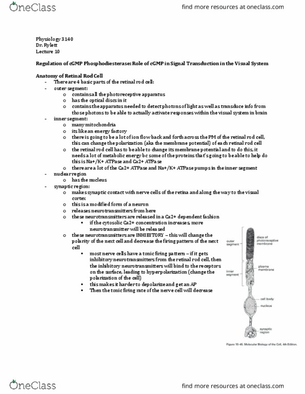
Anatomy of Retinal Rod Cell
- Outer segment - photoreceptive apparatus
o Receives photons of light and initiates transduction of information
o Contains: optical disks + apparatus needed to detect photons of light and
transduce information from photons of light to activate responses in the visual
system of the brain
- Inner segment - many mitochondria
o Energy factor
o Ion flow (Na, K, Ca) back and forth across the plasma membrane of the retinal
rod cell can change the membrane potential of the retinal rod cells
▪ The retinal rod cell has to be able to change its membrane potential
• Become depolarized, repolarized, hyperpolarized etc.
▪ Must be able to move up & down scale of membrane potential
▪ The cell needs a lot of metabolic energy to do that due to the proteins
that accomplish it:
• Sodium potassium ATPase – pump K+ and Na+ across the PM to
rectify the membrane potential
• Calcium ATPase
• Therefore, a lot of ATP is going to be used
o There are lot of calcium ATPase pumps and sodium potassium ATPase pumps
- Nuclear region; Has a nucleus
- Synaptic region - makes synaptic contact with nerve cells of the retina (and along the way to the
visual cortex)
o Modified form of neuron or nerve cell
o Releases neurotransmitters
▪ Note; the neurotransmitters that are released in a calcium dependent fashion
▪ If cytosolic calcium concentration goes up in the cell, it induces release of the
neurotransmitter
• Resting calcium levels in the cell cell induced to have a change in
cytosolic calcium calcium dependent phenomena in cell takes place e.g.
release of neurotransmitters
▪ Neurotransmitter release is calcium dependent event
o Neurotransmitters released are inhibitory
▪ Changes the polarity of the cell & decrease the firing pattern
o Most nerve cells have a tonic firing pattern; input from retinal rod cells & gets more of the
inhibitory neurotransmitter released. The inhibitory neurotransmitter binds to surface
receptor & lead to hyperpolarization (changing polarization of cell) making it harder to
depolarize to get an action potential from the cell. The tonic firing rate of the cell goes down
o MORE RELEASE OF INHIBITORY NEUROTRANSMITTER = LESS INFORMATION GETS SENT
TO THE BRAIN
find more resources at oneclass.com
find more resources at oneclass.com

Responses to Light and Dark Conditions
- In the dark:
o Rod cell is strongly depolarized
o Cation (Na+ and Ca++) voltage gated Ca++ channels open
▪ A lot of sodium and calcium are moving into the retinal rod cells
▪ Membrane potential is depolarized
o Cytosolic Ca++ level high with steady released of transmitter
▪ Get a high rate of transmitter release (inhibitory transmitter)
▪ NOT SENDING ANY INFORMATION TO NEXT NEURON IN LINE, AND NONE TO THE
VISUAL CORTEX (due to the neurotransmitter being inhibitory)
o Rhodopsin is INACTIVE
o No photons hitting rhodopsin = cation channels open and gated = cations (Na, Ca) move
down electrochemical gradient (positive negative, high low)= retinal rod cell
depolarized relative to resting potential = cytosolic calcium increases (higher relative to
resting)
- Light causes:
o Cation channels to close
o Cell hyperpolarizes
▪ Na/K ATPase in inner segment work to re-establish resting levels (Na+ out, K+ in)
o Decreased Ca++ influx and decreased release of transmitter
▪ Ca pumped out by ATPase + channels closed (Ca not going back in)
o Photons of light hit the rhodopsin & it becomes activated through the conformational change
of the retinal chromophore sodium + calcium channels begin closing
o Retina rod cell can send information to the next neuron
- Na+ channels in the image are actually BOTH sodium and calcium channels = selective cation
channels
- Rhodopsin is the GPCR that is going to detect the photons of light
o Portion of the receptor analogous to ligand binding site, is an adapted form of vitamin A
(retinal)
▪ The retinal chromophore can detect photons of light
▪ When photons of light hit the retinal rod cells, it changes the energy state of the
retinal = analogous to a hormone binding to ligand site on GPCR
▪ Photon analogous to ligand
-
find more resources at oneclass.com
find more resources at oneclass.com

Summary
DARK
LIGHT
Membrane potential
Depolarized
Hyperpolarized
Cation channels
Open
Closed
Transmitter release
Increased release
Decreased release
Cytosolic calcium
Higher level
Lower level
Transmitter inhibits postsynaptic neurons
- Illumination frees neurons from inhibition and thus excites them
o Light frees the next nerve cells that the rod cells synapses on in order to communicate
o Taking away inhibitory input to the next nerve cell
o It frees the next neuron to allow it to increase its tonic firing right – equivalent to having an
excitation
- Rate of transmitter release from rod cells is graded to light intensity
o The more light you get = more effect it has
o We are capable of detecting differences in light over a broad range. For this to happen, our
visual system at this level (level of retinal rod cells – where photons are detected) has to be
able to respond to a wide range
o Few photons of light – get few rhodopsin receptors activated
o More photons of light – get more receptors activated
▪ Note: this plays out in the amount of neurotransmitter that is released
o Whole system is synced to the number of photons of light that activate the retinal rod cells
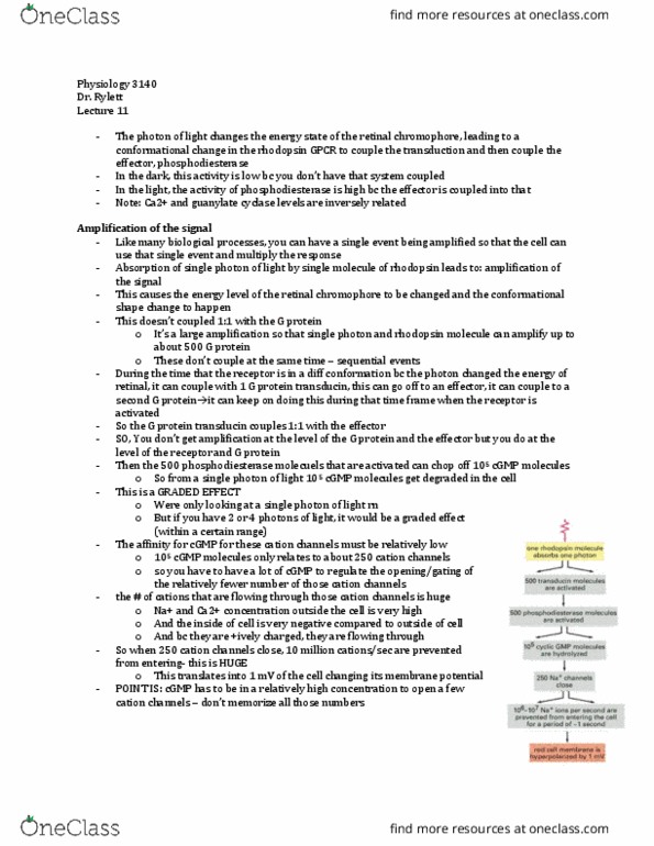
Rod cells contain visual pigment rhodopsin
- Rhodopsin has a light absorbing portion of complex retinal (vitamin A):
o Absorption of light causes conformational change in retinal and cascade of events involving
cGMP
▪ Coupled to G protein and effector
▪ Effector = cGMP dependent phosphodiesterase
• Starts to brings in cGMP
o This is only light –dependent step in vision
- Rhodopsin is the portion of the retinal rod cell that detects light through the chromophore, retinal
(form of vitamin A) that absorbs the photon of light
- Light is absorbed by the chromophore (retinal – portion of the GPCR, rhodopsin) couples G
protein coupling effector (phosphodiesterase) that brings in cGMP
- Note; this is a graded effect
- For example: have a defect where you are deficient in retinal (vitamin A)
o The visual system does not have the chromophore associated with rhodopsin
o Would not be able to detect light
o Would not be able to detect differences in light
o ONLY LIGHT DEPENDENT STEP*
find more resources at oneclass.com
find more resources at oneclass.com
Document Summary
Regulation of cgmp phosphodiesterase: role of cgmp in signal transduction in the. Retinal rod cell where cgmp and cytosolic guanylate cyclase play a key role in regulating our ability to transduce light photons into signals that we can use as information. Inner segment - many mitochondria: energy factor. The inhibitory neurotransmitter binds to surface receptor & lead to hyperpolarization (changing polarization of cell) making it harder to depolarize to get an action potential from the cell. The tonic firing rate of the cell goes down: more release of inhibitory neurotransmitter = less information gets sent. Na+ channels in the image are actually both sodium and calcium channels = selective cation channels. Illumination frees neurons from inhibition and thus excites them excitation. Rhodopsin has a light absorbing portion of complex retinal (vitamin a): absorption of light causes conformational change in retinal and cascade of events involving cgmp, coupled to g protein and effector, effector = cgmp dependent phosphodiesterase.