BI276 Lecture Notes - Lecture 4: Transmission Electron Microscopy, Scanning Electron Microscope, Dark Field Microscopy

Document Summary
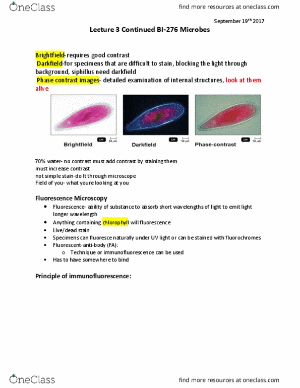
Staining increases contrast at least 70% of bacteria are transparent: observes them while they are not alive. Phase-contrast allows us to view them while they are alive. Dark field microscopy blocks light through the background, shows up on a black background: syphilis needs this to be visible. Uses 2 beams of light, each split by separate prisms. Differences in refractive indexes generates contrast in the image. Fluorescence: the ability of substances to absorb shorter wavelengths of light and emit light of longer wavelengths. Specimens can fluoresce naturally under uv light, or can be stained with fluorochromes: anything with chlorophyll will naturally fluoresce. Required to visualize structures smaller than 0. 2m. Beam of electrons has 100,000 times smaller wavelengths than light. Transmission electron microscopy: magnifies objects 10,000 to 100,000 times, requires sectioning of specimen. Scanning electron microscopy: scans specimens with electron beam and magnifies 1,000 to 10,000 times. Typically use a heat-fixed smear: stick bacteria onto slide before staining.