BIOL 2021 Lecture Notes - Lecture 2: Scanning Electron Microscope, Lipid Bilayer, Sphingosine

13 Jan 2017
School
Department
Course
Professor
Document Summary
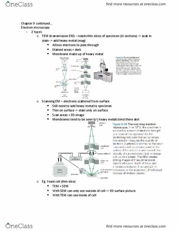
With tem you have e- beam passing through specimen. Specimen is stained with e- dense material. Dark-stained, dense regions regions of reduced e- flux. Scanning electron microscope directly produces an image of the 3d structure of the surface of a specimen. Uses e- that are scattered from specimen"s surface. Specimen is fixed, dried & coated w/ thin layer of heavy metal. Scanned w/ a very narrow beam of e- Usually used to study whole cells or tissue vs. subcellular organelles. Video: the inner life of the cell: leukocytes rolling on endothelial surface, (a bunch of other processes in animation) Video: protein packing: proteins aggregate into clumps, kinase attaches to vesicles w/ proteins embedded in bilayer membrane, other end of kinase attaches to a microtubule & carries the vesicle along it. Functions: barrier to water-soluble molecules, separates inside from outside, cell vs. environment, organelle vs. cytosol, maintains ion gradients, signalling plasma membrane receptors; extracellular signals to intra.