KINE 2031 Lecture Notes - Lecture 4: Cervical Vertebrae, Sagittal Suture, Coronal Suture

11 Dec 2017
School
Department
Course
Professor
Document Summary
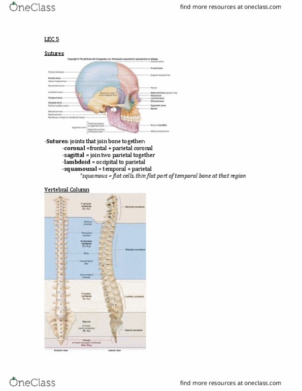
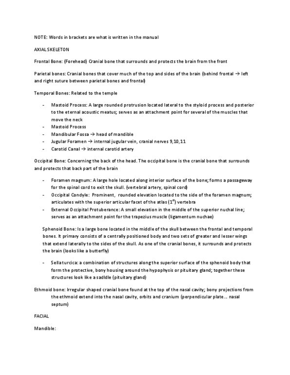
Skeleton divided into axial portion and appendicular portion: axial is the mid of the body, with the appendicular being everything that stems o of it. Skull: frontal bone, at the front of the cranial cavity, behind the frontal bone we have the parietal bones, temporal bone is below the parietal bone. There is an opening in it for the carotid canal. There is an opening between the temporal bone and the occipital bone called the jugular foramen. It is the large vein that drains veinous blood from the cranial cavity out: the mandibular fossa is the depression in the temporal bone, the occipital bone is behind the parietal lobe. It has a large hole that lets the spinal chord come out: on the back of the head there is a distinct bump thats called the external occipital protrudence. The hard part of the roof of the mouth is made of the: maxilla, palatine bone.