PSYC BC 1119y Lecture Notes - Lecture 13: Cyclic Guanosine Monophosphate, Amacrine Cell, Ciliary Muscle

4 Nov 2015
School
Department
Course
Professor
Document Summary
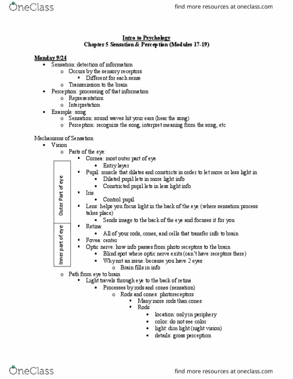
Visible light has a range in wavelength from 400nm (blue) to 700 nm (red) anatomy of the eye. Cornea: stiff part that makes up outside of eye. Ciliary muscles: attached to the lens- change the shape of the lens so we can focus light. Pupil: open hole- smaller lets less light in, larger lets more light in iris: controls the pupil. Pupil and iris control the amount of light that enters the eye. Retina: in the back- light (emr) converted into nerve impulse. Second farthest back layer are the photoreceptors- on the front line of beginning to convert light into electrochemical info. Rods: pick up info under dim light- give us little detail- peripheral vision. Cones: pick up info under good lighting- give us color vision and pick up more detail. Then come the bipolar cells: interneurons that receive glutamate projections from rods + cones.