BCHM-3050 Lecture Notes - Lecture 8: Van Der Waals Strain, Bohr Effect, Amine

26 Jun 2018
School
Department
Course
Professor

Chapter 7: Protein Function and Evolution
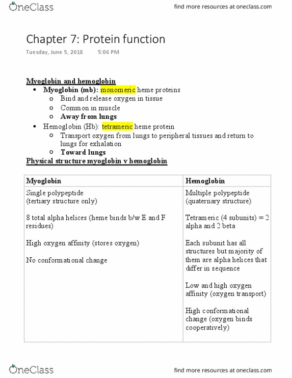
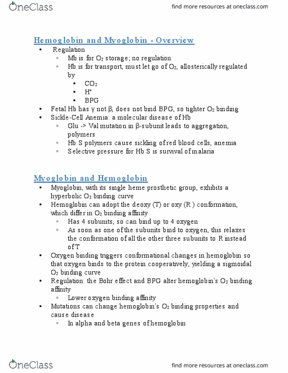
Myoglobin and Hemoglobin
Myoglobin (Mb) – monomeric heme protein that binds and releases oxygen in tissues. Approximately
2mg/g of human muscle tissue for efficient delivery of oxygen to mitochondria.
oMammals have 10-30 fold more myoglobin because they need more oxygen storage to have
them breathe underwater.
oSingle polypeptide (3’ structure ONLY)
oHigher Oxygen affinity than hemoglobin and stores Oxygen
o75% of the alpha helix is globular. Helices are labeled A-H and HEME BINDS BETWEEN E-F
HELICES***
oNO CONFORMATIONAL CHANGE – hyperbolic oxygen binding
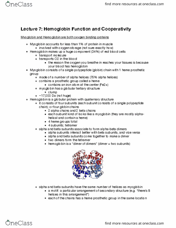
Hemoglobin (Hb) – tetrameric heme protein that transports oxygen from lungs to the peripheral tissues
and returns the CO2 to lungs for exhalation.
oHemoglobin is transported in the vasculature in the RBCs.
oMultiple polypeptides (4’ structure)
oHas 2 copies of alpha subunits and 2 copies of B subunits (why its tetramer)
oEach subunit has THE SAME general structure (1’,2’,3’, or 4’) with some specific differences
in the sequence.
oLow and High oxygen affinity so it can transport oxygen
oHAS CONFORMATIONAL CHANGE – sigmoidal oxygen binding
Heme
Is a derivative of a porphyrin (tetrapyrrole ring system)
In Mb/Hb, the Iron II ion is bound to the protoporphyrin IX via 4 nitrogens.
oCan also use Cu, Mn, and Co ions.
Myoglobin/hemoglobin without heme is called apoprotein or a holoprotein
oHb and Mb with oxygen = oxymyoglobin/oxyhemoglobin
oHb and Mb without oxygen = deoxymyoglobin/deoxyhemoglobin
O2 Binds at Heme
Fe2+ has 6 coordinating positions – 4 nitrogens from the porphyrin.
o1 is a proximal histidine that is on the F8 His.
oThe sixth position is where 1 oxygen binds via a distal histidine called E7 His.
When the iron is in the -deoxy state (no heme or oxygen bound) the proximal histidine (F7) will pull the
iron out of the plane of the porphyrin ring. The porphyrin ring is bent and the iron is being pulled out of
the ring.
When oxygen binds, the iron pops back into the plane, and the ring flattens back out when you form the
–oxy state.
Analysis of O2 Binding
Hemoglobin is in the TAUT state when the iron is in the center, aka being popped out of the ring (bent).
oThe R state (relaxed) is where there is high oxygen affinity.
oA blood oxygen transport protein with a hyperbolic binding curve has a REDUCED O2
DELIVERY vs. pa protein with a sigmoidal binding curve.
find more resources at oneclass.com
find more resources at oneclass.com
Document Summary
Myoglobin (mb) monomeric heme protein that binds and releases oxygen in tissues. Helices are labeled a-h and heme binds between e-f. Helices**: no conformational change hyperbolic oxygen binding. Is a derivative of a porphyrin (tetrapyrrole ring system) In mb/hb, the iron ii ion is bound to the protoporphyrin ix via 4 nitrogens: can also use cu, mn, and co ions. Myoglobin/hemoglobin without heme is called apoprotein or a holoprotein: hb and mb with oxygen = oxymyoglobin/oxyhemoglobin, hb and mb without oxygen = deoxymyoglobin/deoxyhemoglobin. Fe2+ has 6 coordinating positions 4 nitrogens from the porphyrin: 1 is a proximal histidine that is on the f8 his, the sixth position is where 1 oxygen binds via a distal histidine called e7 his. When the iron is in the -deoxy state (no heme or oxygen bound) the proximal histidine (f7) will pull the iron out of the plane of the porphyrin ring.