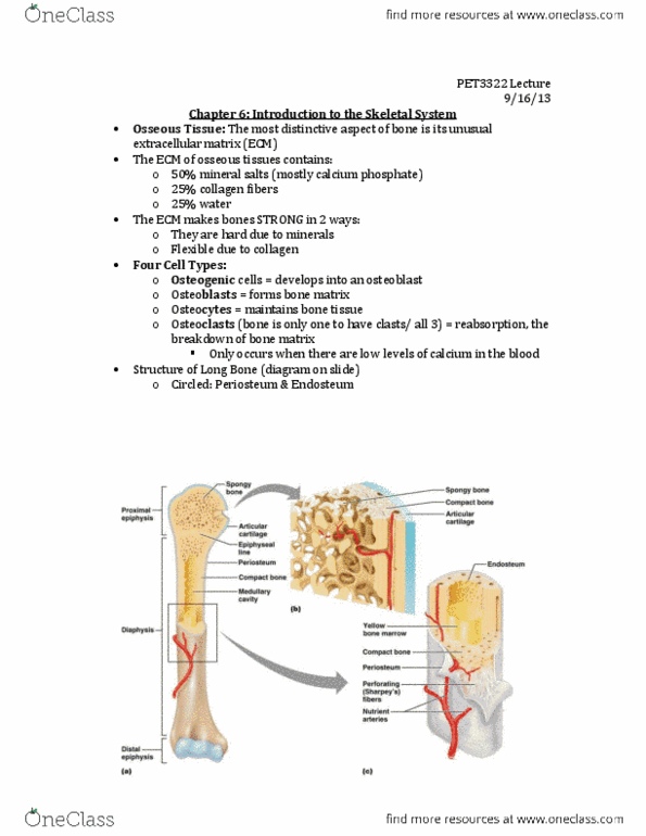
PET-3322 Lecture Notes - Lecture 4: Nutrient Canal, Osteoid, Osteoclast

10 Oct 2017
School
Department
Course
Professor
Document Summary
Skeletal cartilages: the human skeleton initially consists of just cartilage, which is replaced by bone except in areas requiring flexibility. Basic structures, types, and locations: skeletal cartilage: made of highly resilient, molded cartilage tissue that consists primarily of water. Contains no blood vessels or nerves: perichondrium: layer of dense connective tissue surrounding cartilage like a girdle. Contains blood vessels for nutrient delivery to cartilage. Cartilage made up of chondrocytes: cartilage is made up of chondrocytes, cells encased in small cavities (lacunae) within jelly-like extracellular matrix, three types of cartilage: Most abundant type; contains collagen fibers only. Articular (joints), costal (ribs), respiratory (larynx), nasal cartilage (nose tip) Similar to hyaline cartilage, but contains elastic fibers. Thick collagen fibers: has great tensile strength. Growth of cartilage: cartilage grows in two ways: Cartilage-forming cells in perichondrium secrete matrix against external face of existing cartilage. New matrix laid down on surface of cartilage.