MICRO 302 Lecture Notes - Lecture 8: Mycobacterium Tuberculosis, Gram-Negative Bacteria, Gram-Positive Bacteria

28 Jun 2017
School
Department
Course
Professor
Document Summary
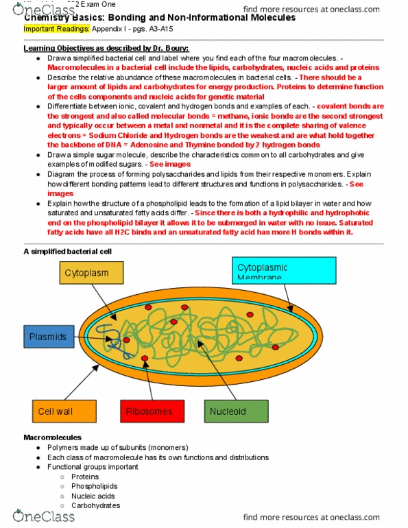
Cell walls of gram positive, gram negative, mycobacterial and archaeal cells. Draw a small segment of peptidoglycan, indicating where and how crosslinking occur and unique structures found within peptidoglycan. Compare and contrast the gram positive, gram negative, archaeal and mycobacterial cell envelopes. Gram positive stain purple and have a double peptidoglycan layer (absorbs more stain). Gram negative have lipopolysaccharides and stain pink because they have a single layer and do not absorb as much stain. Mycobacterial cell envelopes are organized with an inside and outside membrane made of lipids and sugars. Archaeal cell envelopes are very structured and strong and composed of glycerol-ether bond lipids. Explain how subcellular location is important to protein function. Protein subcellular location is important for determination of protein function. Locations work as information about a protein including its protein sequence of amino acids. This is to help creating cellular tools that can accurately predict the outcome of protein targeting in cells.