BIOL 3410 Lecture Notes - Lecture 11: Fibril, Eosin, Micrograph

Document Summary
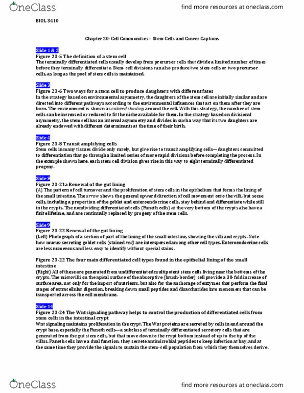
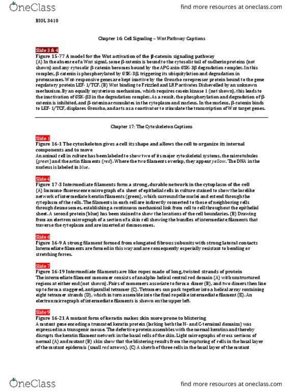
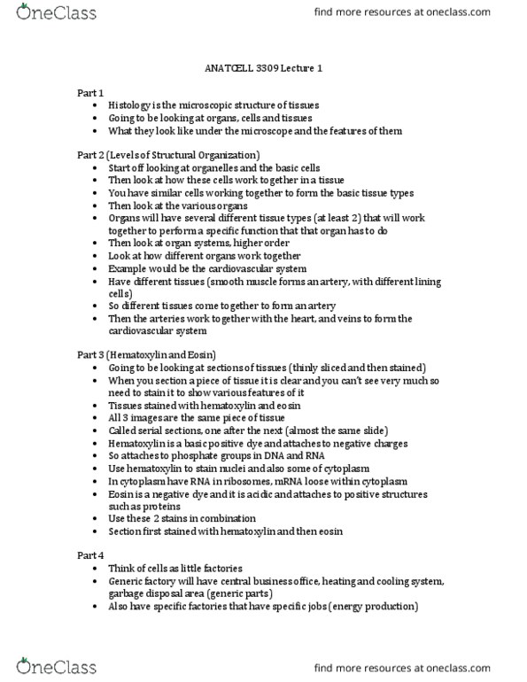
Figure 20-1 multicellular organisms are built from organized collections of cells. This section of cells in the urine-collecting ducts of the kidney was stained with a combination of dyes, hematoxylin and eosin, commonly used in histology. Each duct is made of closely packed (cid:498)principal(cid:499) cells (with nuclei stained red), which form an epithelial tube, seen here in cross section as a ring. The ducts are embedded in an extracellular matrix, stained purple and populated by other types of cells. Simplified drawing of a cross section through part of the wall of the intestine of a mammal. This long, tubelike organ is constructed from epithelial tissues (red), connective tissues (green), and muscle tissues (yellow). Each tissues is an organized assembly of cells, held together by cell-cell adhesions, extracellular matrix, or both. Figure 20-9 collagen fibrils are organized into bundles. The drawings show the steps of collagen assembly, from individual polypeptide chains to triple-stranded collagen molecules, then to fibrils and, finally, fibers.