NUR 306 Lecture Notes - Lecture 9: Superficial Temporal Artery, Cervical Lymph Nodes, Posterior Triangle Of The Neck

Document Summary
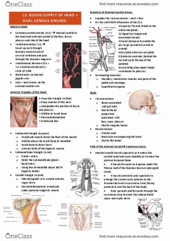
Mandibular joint: joint of the jaw, you would assess the joint for tmj. Vascular: superficial temporal artery (arises from the carotid artery. Carotid artery: lies beneath the sternocleidomastoid muscle. This includes the mandible above, the sternocleidomastoid(laterally), and the midline(medially) The sternocleidomastoid muscle separates the anterior and posterior triangles. The hyoid bone -u- shaped bone, sits below the base of the tongue and the larynx. The tracheal cartilage the main cartilage and the largest cartilage of the trachea. The thyroid gland: thyroid is butterfly shaped, when identifying thyroid first locate the cricoid cartilage, the thyroid lies below the cricoid. In the shaped of a butterfly: two lobes connected by an isthmus. The two lobes lie laterally and wrap around the posterior of the trachea and the esophagus. Should be centrally located in the neck. If you can palpate them the should be less than 1cm, round, smooth, and non-tender.