01:119:116 Lecture Notes - Lecture 2: Crystal Violet, Gram Staining, Plasmolysis

16 Oct 2018
School
Department
Course
Professor
Document Summary
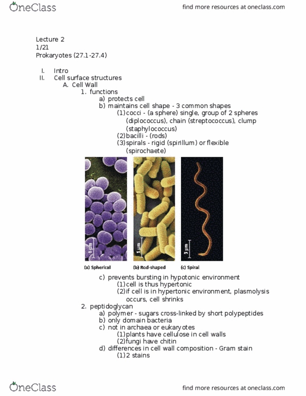
Unicellular; small/microscopic (0. 5 to 5 micrometers in diameter) Three common shapes fig 27. 2: spherical (cid:498)cocci(cid:499, rod-shaped (cid:498)bacilli(cid:499, spiral (cid:498)spirochetes(cid:499, function. Cell wall: maintains cell shape, protects cell, prevents bursting (in hypotonic environment lower concentration. 1/24/13: complex w/ outer membrane made of lipopolysaccharides, lipopolysaccharides could be toxic to host, less peptidoglycan, more resistant to bacteria. In gram staining, the dye will rinse out with alcohol: cell wall surrounded by sticky polysaccharides or proteins, facilitate attachment to substrate or other cells, well defined layer called capsule. Fig 27. 4: not so well defined layer called slime layer. Some species have fimbriae which are hair-like appendages that allow cells to stick to substrate or other cells fig 27. 6. Pili are appendages that pulls cells together before dna transfer occurs between cells: sex pili. Fig 27. 7: eukaryotic flagella has outer plasma membrane, prokaryotic flagella moves by using pumping proton ions out and in causing to spin this rod to motor the movement.