01:447:380 Lecture Notes - Lecture 11: G Banding, Allosome, Microscope Slide

27 Apr 2018
School
Department
Course
Professor

LECTURE 11
Cytogenetics
Techniques For Chromosome Analysis
Numerical And Structural Chromosome Abnormalities
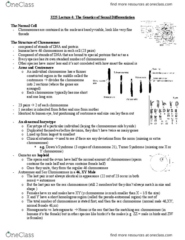
I. Human Chromosome info
a. 23 pairs
i. 22 pairs autosomes : 1-22
1. 1 = largest autosome
2. 22 = smallest
ii. 1 pair = sex chromosomes : XX = female, XY = male
b. Karyotype
i. 2 contexts:
1. Idiidual’s hroosoe status : HOW MANY & WHICH “EX
CHROMOSOMES & ABNORMALITIES?
2. Piture of the idiidual’s atual hroosoes
ii. A karyotype contains three elements, separated by commas:
1. Total number of chromosomes
2. Sex chromosomes
3. Any abnormalities
iii. Normal female = 46,XX Normal male = 46,XY
1. Male with Down syndrome (has extra chromosome 21) = 47,XY,+21
2. Female with Turner syndrome (missing an X chromosome) = 45,X
3. Male missing one chromosome 6 = 45,XY,-6
iv. G-banding gives each chromosome arm a unique combination of black, white
and grey bands
1. Cells are gro i ulture, ad speial addities are used to halt a ell’s
mitosis in metaphase, when the chromosomes are most condensed and
easiest to see under a microscope
2. Culture is dropped onto a glass microscope slide, whereupon the cells
burst open and the chromosomes spread out (a metaphase spread)
3. The chromosomes are treated with enzymes that partially digest the
proteins in the chromatin, then dyed with Giemsa (a dark blue-black
dye), hih is hat gies the proess te ae G adig
4. A-T rich regions stain more strongly than G-C rich regions
5. Each chromosome arm has a unique pattern of thick and thin black, grey
and white bands
6. G-banding also provides a system for localizing genes on their
chromosomes
a. For each chromosome arm, the most centromeric band = 11.1;
numbers increase as you go toward the telomere
7.
find more resources at oneclass.com
find more resources at oneclass.com


.jpg)
