01:830:301 Lecture Notes - Lecture 10: Optic Chiasm, Visual Cortex, Receptive Field

Document Summary
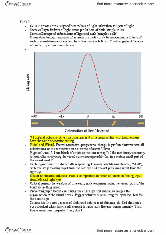
Temporal half of retina > ipsilateral visual cortex. Nasal half of retina > contralateral visual cortex. Left visual fields (both eyes) > right visual cortex. Right visual fields (both eyes) > left visual cortex. Primary visual cortex - also known as: area v1. The fovea receives a large representation within this cortex. Cortical magnification: 80% of cells devoted to central 10 degrees. 6 layers: inputs from lgn received in layer 4. Simple cells respond to edges and bars of specific orientations. Elongated receptor fields with clearly-demarcated on and off regions. Because of such a property, the exact positioning of edge/bar not required. First site of binocular cells, moving from eye to the brain. These cells have two receptive fields (one in the left eye and one in the right eye), to account for and respond to stimuli obtained from both eyes. Ocular dominance - slightly stronger responses to one eye.