BIOL 320 Lecture Notes - Lecture 9: Exocrine Gland, Stratified Squamous Epithelium, Simple Squamous Epithelium

14 Jan 2017
School
Department
Course
Professor
Document Summary
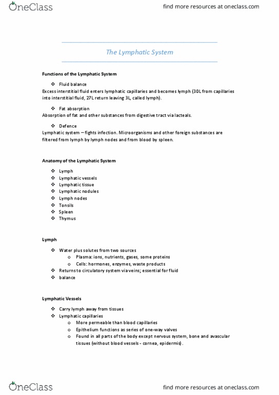
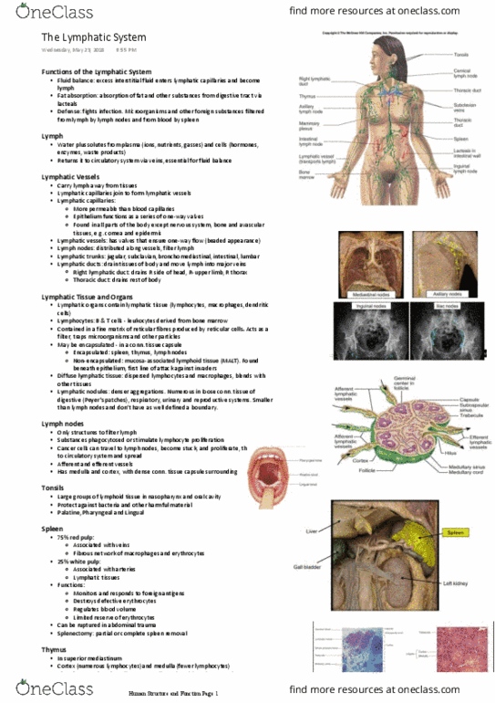
Lymphatic, digestive, respiratory, urinary, and reproductive system histology. Returns fluids that have leaked from vessels back into blood. Bean shaped lymphatic organs connected by vast network of lymphatic vessels. High concentration in the upper limbs, axillary and cervical regions. Entraps and destroys old erythrocytes and platelets. Left upper side of the abdominal cavity, below the diaphragm. Capsule surrounds it to protect underlying tissue of red and white pulp. Free and fixed phagocytes remove abnormal red blood cells and other antigens from the blood. It becomes sensitized to them and produces antibodies to counteract them. From sinuses of the red pulp trabecular veins eventually into splenic vein. Appears blue due to lymphocyte nuclei stains. Extracellular fluid that has entered lymphatic vessels. Further components of lymph are added by the lymphatic organs. They eventually make their way to the stratum spinosum of the epidermis. Collections of lymphatic tissue associated with the inside of the throat and mouth.