KINE 318 Lecture Notes - Lecture 2: Anterior Cruciate Ligament Injury, Tensor Fasciae Latae Muscle, Gluteus Medius Muscle

7 Mar 2017
School
Department
Course
Professor
Document Summary
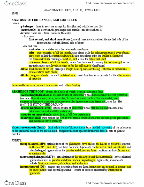
The knee is a ginglymus (hinge joint) allowing flexion and extension (slight medial and lateral rotation). The knee is weak in bony arrangement but this is compensated for through the support of ligaments and musculature. Designed primarily to provide stability in weight bearing and mobility in locomotion. Distal end of femur: articulates with the tibia & patella: has 2 condyles: lateral & medial (larger from front to back, form a hollowed area to receive the patella. Tibia: consists of 2 tuberosities designed to receive the condyles of the femur: tuberosities divided posteriorly by a groove called popliteal notch. Patella: largest sesamoid bone in the body: lies within the tendon of the quadriceps, gives anterior protection to the knee joint. Quadriceps (vastus medialis, vastus lateralis, vastus intermedius, and the rectus: extension, pes anserine: (from distal to proximal): common insertion of the sartorius, gracilis, semitendinosus, on the tibia (medial side) femoris) Anterior thigh muscles: sartorius and quadriceps femoris group.