BIO 201 Lecture Notes - Lecture 4: Sickle-Cell Disease, Fried Egg, Globin

9 Feb 2017
School
Department
Course
Professor
Document Summary
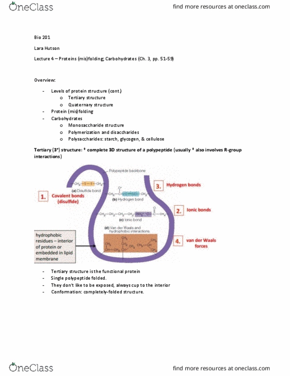
3d structure of a single polypeptide chain resulting from 2 structure + r-group interactions. Complete 3d structure of a polypeptide (usually also involves r-group interactions) Bond strength from strongest to weakest: covalent ionic hydrogen van der. Interior of protein or embedded in lipid membrane. For proteins comprised of a singly polypeptide: Catalyzes degradation of something else (destroys it) Interactions between multiple polypeptide subunits: multi-subunit proteins only. Homomultimers: comprised of 2 or more identical polypeptide subunits. Heteromultimers: comprised of 2 or more different types of polypeptide subunits. Hemoglobin (proteins in red blood cells that carries o2) - has 2 identical (cid:757)-globin + 2 identical (cid:758)-globin subunits + heme group (organic molecule, contains 1 iron ion where o2 binds)) Misfolding can be due to protein production or gene mutation. Amino acid change due to error in protein production or gene mutation. Common in regions where malaria is widespread. Cells get stuck in blood vessels and can"t carry blood.