BMB 401 Lecture Notes - Lecture 3: Hemoglobin, Oligomer, Ramachandran Plot

16 Dec 2016
School
Department
Course
Professor
Document Summary
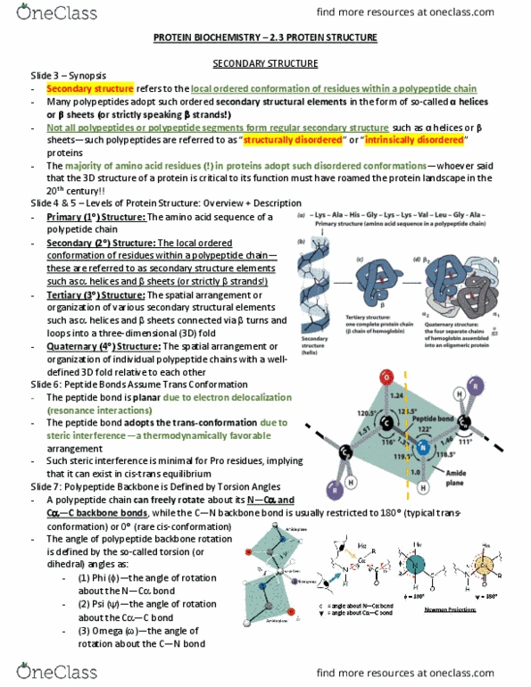
1 = linear amino acid sequence of a polypeptide. 2 = local ordered conformation of residues within a polypeptide chain. These secondary structure elements include a helices and b sheets (strands!) 3 = spatial arrangement of these secondary structures like the a helices and b sheets. They can be connected via b turns and loops into a 3d fold. 4 = spatial arrangement of individual chains when they come together to form a well-defined 3d structure. Not all proteins experience all 4 levels necessarily. A small protein may only be comprised of one pol(cid:455)peptide a(cid:374)d the(cid:396)efo(cid:396)e does(cid:374)"t ha(cid:448)e a (cid:395)uate(cid:396)(cid:374)a(cid:396)(cid:455) st(cid:396)u(cid:272)tu(cid:396)e fo(cid:396) e(cid:454)a(cid:373)ple. The bond itself is planar due to resonance interactions and it adopts this trans conformation because it reduces steric interference and therefore is thermodynamically favorable. The exception however is for proline which is the only fully cyclic amino acid. In this case steric interference is minimal so it can exist in a cis-trans e(cid:395)uili(cid:271)(cid:396)iu(cid:373)!