BIS 104 Lecture Notes - Lecture 3: Cell Fractionation, Cell Membrane, Immunoprecipitation
27 Jan 2018
School
Department
Course
Professor
Document Summary
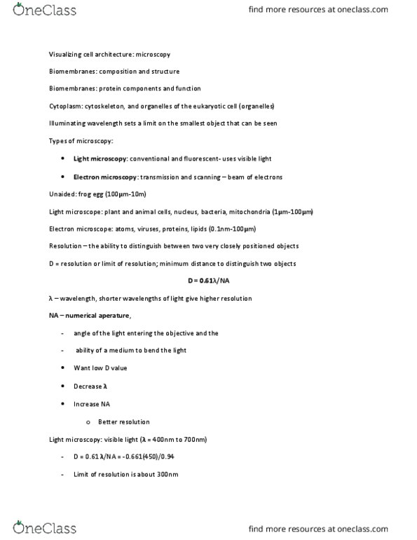
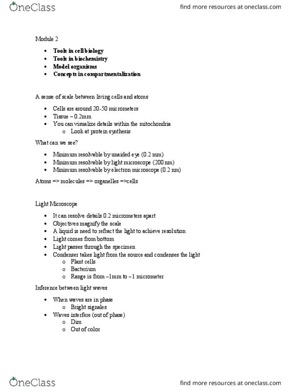
Transmitted light microscopy: d = 0. 61(lamda)/ n sin (alpha) Lamda = wavelength of light ~ 450nm. Alpha = angle that light enters objective. Maximum = sin 90 = 1: practical limit: (0. 61)(450) / (1. 4) (1) = 200nm. Fluorescence microscopy: fluorophore fluorescent molecule. Emits photon of lower energy (higher lamda: indirect immunofluorescence. Add secondary antibody that requires primary antibody. Secondary antibody has fluorophore: live fluorescent imaging. Take gene to gfp fuse to gene of interest. Electron microscopy: electrons have shorter wavelength than light. Down to 0. 03nm = distance: can see lipid bilayer, electrons can be accelerated by high voltage, problem. Cells must be fixed no gfp imaging. Samples must be sliced thin low contrast general staining. Core concepts of cell biology localization of protein can tell something about their function: cell fractionation indirect immunofluorescence, gfp localization. Proteins work in complex and network: immunoprecipitation. Cell processes are dynamic: cyclical cell cycle, plasticity ability to turn on and off.