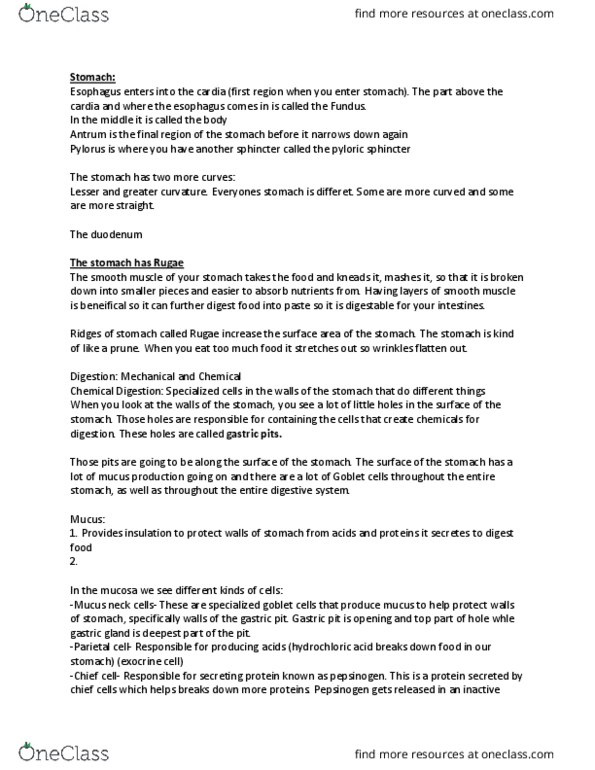
ANATOMY 201 Lecture Notes - Lecture 9: Intestinal Gland, Pylorus, Circular Folds

22 May 2018
School
Department
Course
Professor

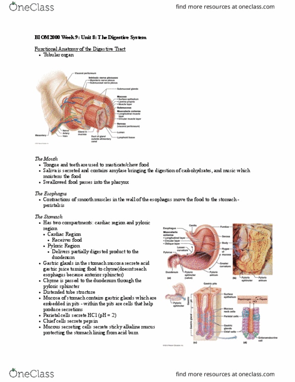
The Lower Digestive Tract: Small and large intestines
Small Intestines:
-From mouth to bottom of stomach is about 50 cm
-750 cm length of small intestine
-Large intestine is only about 4 ft or 5 ft long
-Upper digestion is 2.5 feet long
Organization:
Small intestine divided into three major parts:
A. Duodenum: First region of small intestine that you hit as you leave the
stomach. Once you go through pyloric sphincter you enter small intestine,
specifically the duodenum.
B. Jejunum: After Duodenum
C. Ilium: Closest to stomach vs furthest away from stomach
Ends at the iliosecal valve- A sphincter that divides the small intestine from the
large intestine.
Structures:
Primary function of small intestine is nutrient absorption. That paste (food) enters
small intestine where it is being absorbed as nutrients inside your blood.
-A lot of different little ridges called circular folds. Circular folds that are on lumen
side of small intestine serves a purpose to give small intestine a larger surface
area, therefore more absorption of nutrients.
-Inner layer of small intestine are epithelial cells.
-First you have epithelial layer
-The you have submucosa layer
-Then you run into a layer of circular muscles which will help with contractions
and push food along through digestive system so it does not stay in one place for
too long
-Outside of it you have a serosal layer that creates fluid so your intestines can
move around your gut without rubbing against stuff and damaging itself. It
provides lubrication so intestie does’t ru agaist itself.
-Inner side of luminal wall of small intestine you see small structures that are
microscopic. Finger like projections sticking up from wall. The wall in the inside of
your intestines has all these fingers sticking up that gives it a soft appearance.
find more resources at oneclass.com
find more resources at oneclass.com

These fingers are called villi, and are there to serve another purpose- to further
increase the surface area of the small intestine.
-These villi are going to be covering everything. The picture here shows a circular
fold underneath the villi. Circular folds are big and visible to naked eye while villi
are very small
-Villi are made up of epithelial cells. The primary function of epithelial cells are for
absorption. These specific cells we are talking about are Enterocytes which are
really good at absorbing nutrients (fats, proteins, sugars, etc.). The enterocyte has
microvilli which are FURTHER WAYS that the body can increase surface area.
-Three major structures that increase surface area
Circular folds
Villi- finger like projections
Microvilli- Absorbs nutrients
In between the villi are called Crypts. A crypt is going to be like a pit like in the
stomach. The intestinal crypts are similar to pits, they function like glands and a
lot of secretion goes on.
You have a lot of endocrine cells in the crypts to better absorb or absorb less
nutrients from your food.
-Besides enterocytes, we also see goblet cells to lubricate the food so stuff
does’t get stuk to strutures, et.
-Below the surface are crypts
-Paneth cells: Kills stuff before they enter in your bloodstream
Lacteal:
Inside each villus you have a lot of blood flow.
There is fat that the body handles differently with fats- it absorbs them but
does’t put it diretly i lood syste
It enters in the lymphatic system. (Yellow tube is a lymphatic vessel known as a
lacteal, which is a lymphatic vessel whose primary function is to absorb fats and
transport fats to lympathic system for further processing before it enters the
blood)
Enters into a lymph node- takes white blood vessels, cleans it, and spits it out.
Fats get absorbed in lacteal, which is a vessel that transports fat to lymphatic
vessel before it gets in blood.
find more resources at oneclass.com
find more resources at oneclass.com

Histology:
Fingerlike projections: Villi
Grooves in surface going down to tissue are: Crypts
White dots: Goblet cells
On every villi and crypt there are enterocytes that allow for nutrient absorption
Layer of epithelium is known as the mucosal layer
Then you have the submucosa
And then you have smooth muscle layer
And then connective tissue of serous membrane
Celiac Disease
-An allergy to the gluten protein which is found in grains and wheat.
-If you have an allergy to it it can make you sick
-If you eat a lot of gluten and you have celiac disease, gluten will get to small intestines and it
will cause all the cells in your small intestine to get inflamed.
-Everything Is inflamed, a lot of blood flow in there, increased cell death, buildup of scar tissue
over time
-If you are consistently eating wheat and have gluten allergy, you will damage the wall of your
intestines. The finger projections will go away and it will be a mess
-Losing villi over time will decrease the efficiency of nutrient absorption
Large intestine: Short and fat
Three finger widths in diameter
Has a very specific purpose
Major components first-
• Enter large intestine after you are done going into large intestine through ileocecal
valve, which will close off the opening to your large intestine (opening from ilieum, last
part of small intestine, to opening of secum).
• Cecum is large fat part of large intestine and it is a food collecting area. It will fill in and
sit inside the cecum.
• As food enters into the cecum, it just hangs out there for a while
• The cecum is first area you enter, and then after you leave the ceum, you get to the
ascending colon.
• Right angle turn known as the Right colic flexure
• Transverse colon
• Left colic flexure
• Descending colon runs downwards until you run into S shaped curve called sigmoid
colon
• Get to rectum, holding area for poop
find more resources at oneclass.com
find more resources at oneclass.com
Document Summary
The lower digestive tract: small and large intestines. From mouth to bottom of stomach is about 50 cm. Large intestine is only about 4 ft or 5 ft long. Small intestine divided into three major parts: duodenum: first region of small intestine that you hit as you leave the stomach. Once you go through pyloric sphincter you enter small intestine, specifically the duodenum: jejunum: after duodenum, ilium: closest to stomach vs furthest away from stomach. Ends at the iliosecal valve- a sphincter that divides the small intestine from the large intestine. Primary function of small intestine is nutrient absorption. That paste (food) enters small intestine where it is being absorbed as nutrients inside your blood. A lot of different little ridges called circular folds. Circular folds that are on lumen side of small intestine serves a purpose to give small intestine a larger surface area, therefore more absorption of nutrients. Inner layer of small intestine are epithelial cells.