PSYCH 7A Lecture Notes - Lecture 3: Occipital Lobe, Frontal Lobe, Auditory Cortex


19
PSYCH 7A Full Course Notes
Verified Note
19 documents
Document Summary
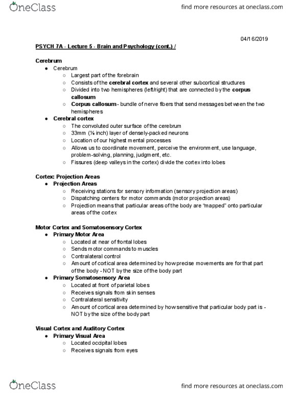
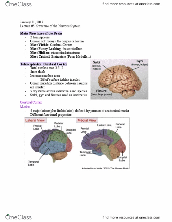
Cerebral cortex: convoluted outer surface of the cerebrum, 3mm ( in. ) layer of densely packed- neurons. Visual cortex and auditory cortex: primary visual area, located occipital lobes, receives signals from eyes, contralateral fxn: left visual field processed in right hemisphere and vice versa. Primary auditory area: located temporal lobes, receives signals from ears, not contralateral- both hemispheres receive signals from both ear. Wernicke"s area and broca"s area: wernicke"s area, located in (usually left) temporal lobe, damage causes wernicke"s aphasia: impaired ability to comprehend. Involved in understanding language speech and to think of words to express one"s thoughts: broca"s area, located in (usually left) frontal lobe, damage causes broca"s aphasia: one is able to understand language, but. Involved in language production speaks slowly and laboriously: angular gyrus, located btwn visual cortex and wernicke"s area, translates visual info into auditory info, damage leads to impairment of reading ability. Visual fields processed contralaterally: right visual field processed in left, vice versa.