BME 80H Lecture Notes - Lecture 17: Cytogenetics, Centromere, Karyotype

11 Jun 2018
School
Department
Course
Professor

17th lecture- Cytogenetics, Chromosomal Aberrations and Human Disease
(chapter 7 p. 152/150; chapter 6 all)
Part I: Cytogenetics: the study of chromosomes and their behavior in a cell.
I. Examining human chromosomes
A. Chromosomes have distinctive banding patterns when treated with
dyes (figures 6.4, 6.8)
- Each arm is subdivided into numbered regions from the centromere to the
telomere
- Within each region, bands are identified by number
- So, you can get an address
- Ex. 1q2.4 means chromosome number (1), the arm (q), the region (2), and
the band (4)
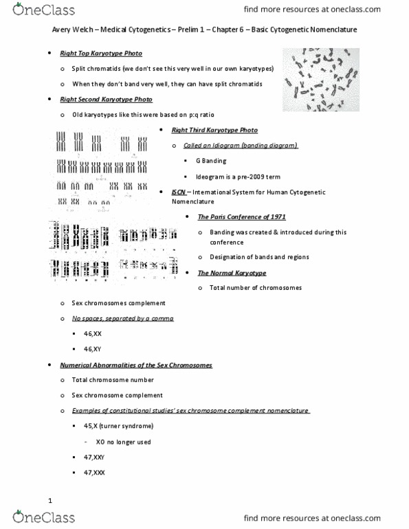
B. Nomenclature of Chromosomes and their parts
Classifications (figures 6.2/6.3, 6.5,
http://www.biologia.uniba.it/rmc/2-YAC-BAC/BAC-
Chromosome/ideograms/10.html):
Whole Chromosome: centromere location,
chromosome size and banding pattern.
Chromosome Arms: Separated by the centromere
p (short) and q (long)
Chromosome Regions: numbered starting at the
centromere
Chromosome Bands: within each region. Numbered
in the direction away from the centromere. (note error
in book figure 6.5)
find more resources at oneclass.com
find more resources at oneclass.com

C. Analyzing karyotypes
1. Visible chromosomal aberrations:
translocation (t)- material from one chromosome is
transferred to another, non-homologous
chromosome.
duplication (dup)- segment of a chromosome is
duplicated.
deletion (del)- segment of a chromosome is deleted.
Nomenclature of Chromosomal aberrations (Table 6.2)
2. Chromosome Painting (figure 6.9)
- Chromosome painting shows the location of translocations, duplications,
deletions, and aneuploidies
II. Diagnosis of chromosomal abnormalities
A. Adults: make a Karyotype using cells from one of the following sources
(figures 6.6, 6.7):
- Draw 10-20 mL of blood
- Add drops of blood and phytohemagglutinin to stimulate mitosis then let it
incubate
- Add Colcemid to culture for 1-2 hours to stop mitosis in metaphase
- Centrifuge to concentrate cells and add a low-salt solution to eliminate blood cells
and swell lymphocytes
- Digitized chromosome images
find more resources at oneclass.com
find more resources at oneclass.com
Document Summary
17th lecture- cytogenetics, chromosomal aberrations and human disease (chapter 7 p. 152/150; chapter 6 all) Part i: cytogenetics: the study of chromosomes and their behavior in a cell: examining human chromosomes, chromosomes have distinctive banding patterns when treated with dyes (figures 6. 4, 6. 8) Each arm is subdivided into numbered regions from the centromere to the telomere. Within each region, bands are identified by number. 1q2. 4 means chromosome number (1), the arm (q), the region (2), and the band (4: nomenclature of chromosomes and their parts. Whole chromosome: centromere location, chromosome size and banding pattern. Chromosome arms: separated by the centromere p (short) and q (long) Nomenclature of chromosomal aberrations (table 6. 2: chromosome painting (figure 6. 9) Chromosome painting shows the location of translocations, duplications, deletions, and aneuploidies. Diagnosis of chromosomal abnormalities: adults: make a karyotype using cells from one of the following sources (figures 6. 6, 6. 7): Add drops of blood and phytohemagglutinin to stimulate mitosis then let it incubate.