MEDT270 Lecture Notes - Lecture 1: Scanning Electron Microscope, Macconkey Agar, Phase-Contrast Microscopy

27 Oct 2016
School
Department
Course
Professor
Document Summary
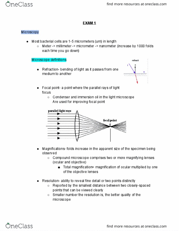
Most bacterial cells are 1-5 micrometers in length. Sizes go in x 1000 folds: largest 1m - 1mm - 1um - 1nm smallest. Refraction bending of light as it passes from one medium to another. Focal point a point where parallel rays of light focus. Folds increase in the apparent size of the specimen being observed. Compound microscope comprises 2 or more magnifying lenses (ocular and objective) Resolution ability to reveal detail or two points distinctly, reported by the smallest distance between two closely-spaced points that can be viewed clearly. The smaller the number of resolution the better quality of the microscope. Gram (+) has very thick peptidoglycan layer, gram (-) is thin. Shapes of cells: cocci small balls can be in clusters, chains, pairs, or tetrads. Arrangement is very important in differentiating bacteria: bacili coccobacilli are very small ovals, almost rod shaped, miscellaneous shapes, or long strings. Arrangement is not important: spirochetes very long spiral shaped string.