BIO 226L Lecture Notes - Lecture 2: Crystal Violet, Gram Staining, Unicellular Organism

Document Summary
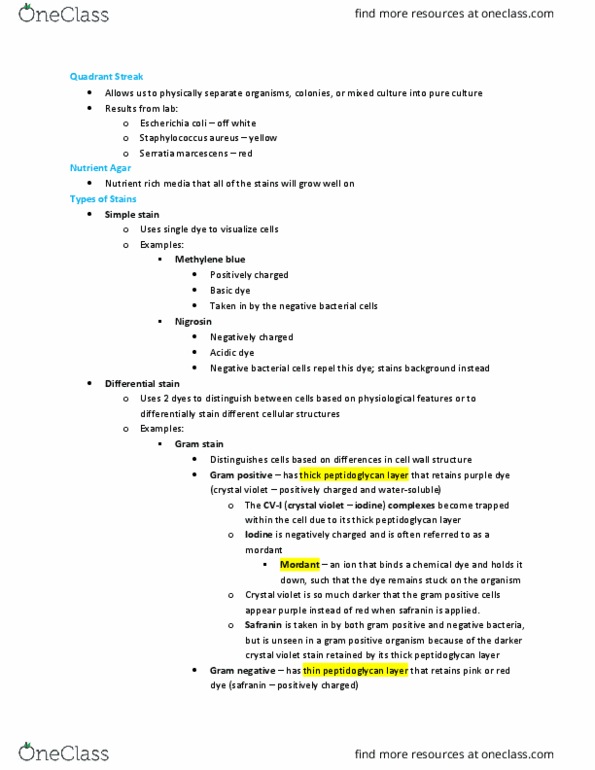
Can survive extreme conditions - nutrients off gases. Modified cellular envelope - strong polysaccharide shells. No peptidoglycan have pseudomurein (polysaccharide nag) Architectural makeup of cellular envelope unique from bacteria inhabit niches that limit bacteria. Gram - staining technique 1884 determine bacteria via staining cell wall. Contrast dye (ionic stain) - crystal violent penetrates the cellular envelope and completely saturates by charge interaction the counter ions in the bacterial cell. Add mordant (iodine) - causes the crystal violet to aggregate into crystal complexes. Both gram - & + are purple at this stage. Alcohol removes the stain (crystal violet aggregates) from gram negative escape through the thin layer of peptidoglycan. Thick layer can"t escape the gram positive. Time is an important factor - too much time and the crystal violet will escape from gram positive as well. Gram + is purple, gram - is transparent. Safranin (counter stain - pink when light passes through) present in both gram positive and negative.