PSYC 406 Lecture Notes - Lecture 4: Retina, Positron Emission Tomography, Hemoglobin

Document Summary
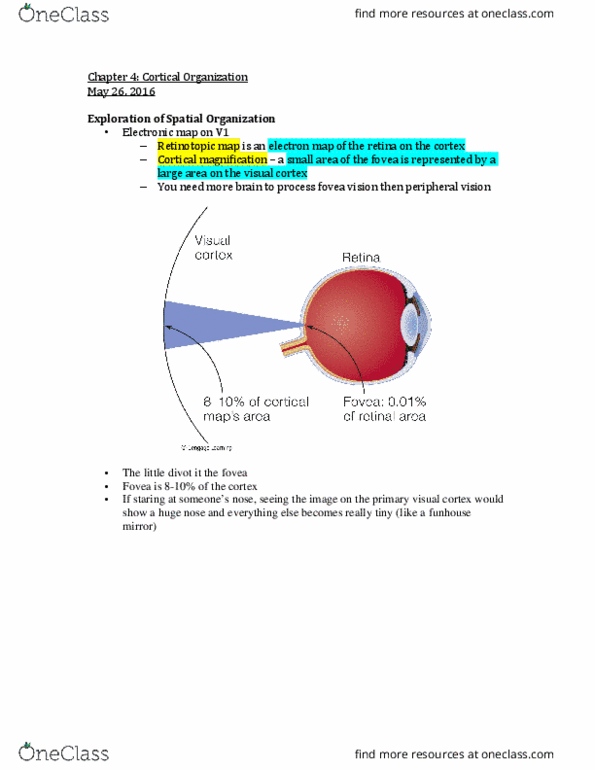
Retinotopic map: electron map of retina on cortex. Cortical magnification: small area of fovea represented by large area on visual cortex. Changes in flow show changes in brain activity. Brain activity determined by subtracting activity in control state from simulation activity. Hemoglobin carries oxygen and contains ferrous molecule, magnetic. Brain activity takes up oxygen, makes hemoglobin more magnetic. Fmri determines activity of areas of brain by detecting changes in magnetic response of hemoglobin. Receptive field at the same location on the retina are within a column. Neurons within fire maximally to the same orientation of stimuli. Adjacent columns change preference in an orderly fashion. 1 mm across cortex represents entire range of orientation. Tiling: columns working together to cover the entire visual field. Specific part of brain is removed or destroyed. Animal restrained to determine which perceptual abilities remain. Results reveal which portions of brain are responsible for specific behaviors. Monkey trained to pick food from cylinder.