LING 390 Lecture Notes - Spin–Lattice Relaxation, Magnetization, Hemoglobin

5 Dec 2012
School
Department
Course
Document Summary
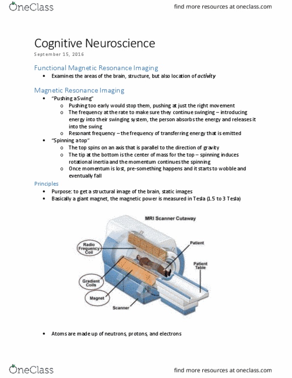
Regular mris just give you structure. functional looks for modules. you need anatomy for functional too. In order to answer this question, we will look at: It"s too difficult to understand how the signal is generated for this class. Fmri - magnetization: magnetic fields and spins (protons in the body become aligned to the field) Fmri -excitation: radio frequency pulses knock protons over - relaxation times: then they go back to their state of alignment with the field, they emit energy that the coil receives. Fmri - tissue contrasts: make it possible to encode the spatial information of signal. Fmri - bold and t2* : concentration of hydrogen in an area (anatomical mri); amount of oxy- vs. deoxyhemoglobin in an area (functional mri) T1 is the time is takes the protons to realign with the magnetic field. T2 is the time it takes for the protons to get back to spinning in unison.