BIOL 1004 Lecture Notes - Venae Cavae, Bronchial Artery, Thoracic Duct

28 Jan 2013
School
Department
Course
Professor
Document Summary
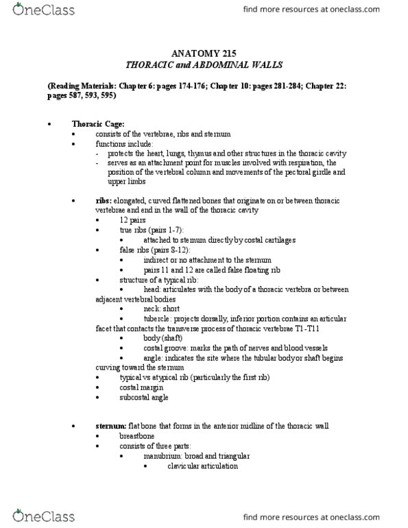
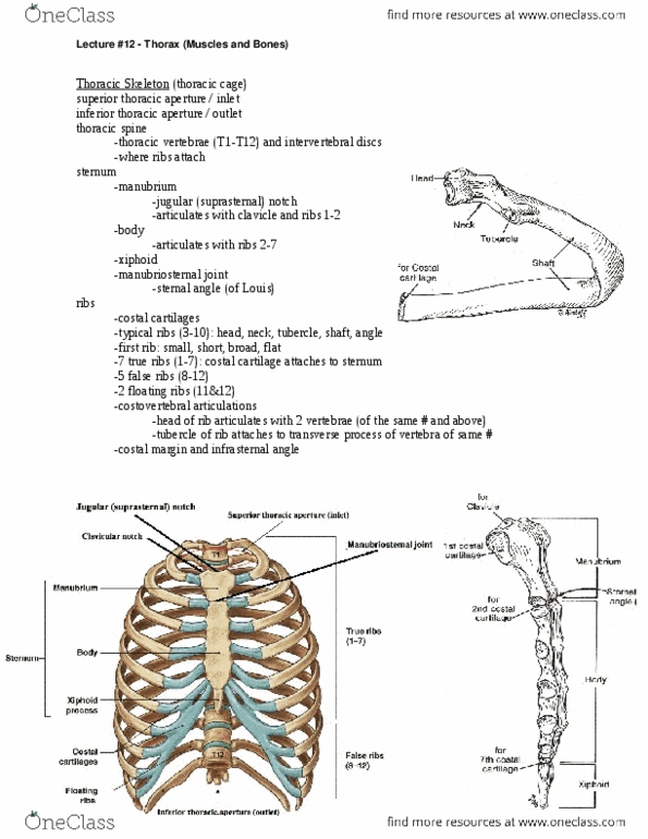
Thorax: it would be way too long to describe every structure. So, this list is really to jog your memory. For each structure, try to recall shape, location, function, and relationships. Check notes to verify your memory: indicates a relationship to another structure. Try to recall what the relationship is: mn. means mnemonic, thoracic wall, thoracic vertebrae (t1-t12) Vertebral foramen ( vertebral canal spinal cord) Sternal angle (more accurately, sternal angle rib 2) Xiphoid process (ossifies later in life: sternum, ribs atypical) innermost. Ribs 1-7 attach to sternum via costal cartilage (1 and 2 are. Ribs 8-10 attach using costal cartilage of superior rib. Ribs 11-12 are floating, no tubercles or necks. Ribs slant inferiorly (e. g. rib 2 (sternal angle) in line with t4/t5) Costal cartilage ( sternum, made of hyaline cartilage) Ic vein azygos/hemizygos, internal thoracic/musculophrenic veins. Ic arteries internal thoracic/musculophrenic, superior intercostals/thoracic aorta arteries. Ic nerves: are ventral rami of t1-t11 spinal nerves: lymphatics.