BIOL303 Study Guide - Paraxial Mesoderm, Lateral Plate Mesoderm, Wnt Signaling Pathway

18 May 2013
School
Department
Course
Professor


Document Summary
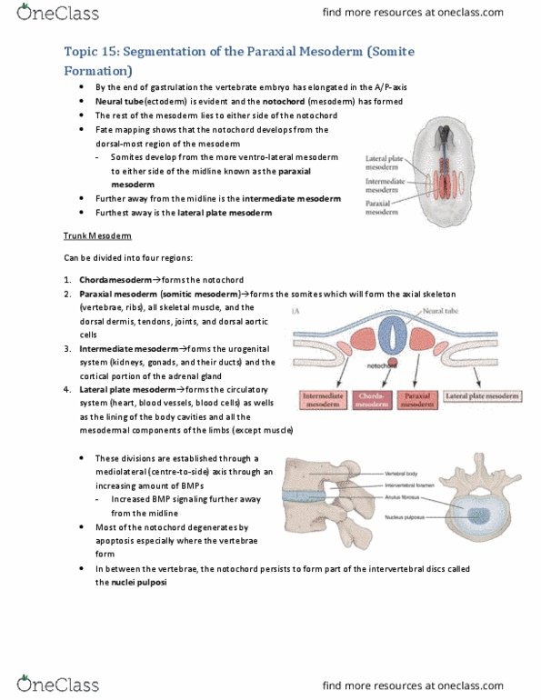
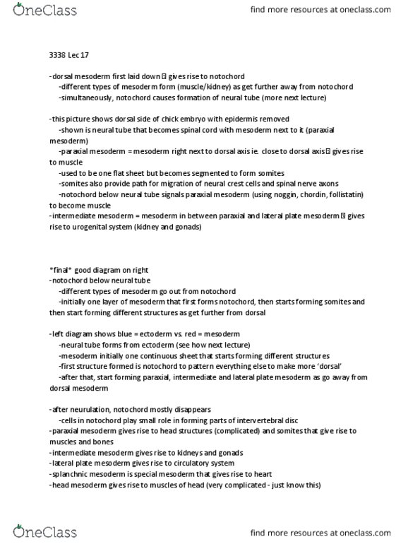
Sclerotome (vertebrae, ribs, rib cartilage) myotome (musculature of back, ribs, limb muscle) dermatome (back dermis) endothelial cells (vascular cells in dorsal aorta) syndotome (tendon) Notch receptor protein, has signal pathway associated with gene expression for boundary formation o. Delta-like3 ligand for notch to initiate signal pathway (juxtacrine signaling) Fgf expression trigger oscillating clock signal to make notch. Ephrin-b2 actual ligand for somite and unsegmented paraxial mesoderm boundary formation. Epha4 (tyrosine kinase) receptor in anterior of unsegmented paraxial mesoderm. Explain the results of the following experiment involving protein notch. Quail presumptive boundary cells transplanted into unsegmented paraxial mesoderm of chick. Quail non-boundary cells transplanted into unsegemented paraxial mesoderm of. Quail non-boundary cells tramsplated into unsegmented paraxial mesoderm of chick followed by local ectopic (unusual place) expression of notch via induction of graft by electroporation to activate notch. Binding cause notch domain to be cleaved and that polypeptide piece goes into the nucleus to influence gene expression (i. e. form gaps b/w segments)