COMM1020 Lecture Notes - Diastole, Purkinje Fibers, Bradycardia

Document Summary
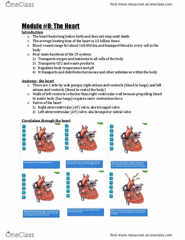
Right ventricle tricuspid valve right atrium pulmonary valve lungs . Pulmonary veins left ventricle bicuspid valve left atrium aortic valve aorta . Superior/inferior vena cava + coronary right ventricle. Histology: cardiomyocyte have desmososmes and gap junctions, intercalated disks, nucleus, sarcolemma and tons of mitochondria. Cardiomyocyte ap: has a much slower repolarization due to a plateau period in which. Ca2+ flows into cell and k+ flows out. Sodium potassium atpase, makes atp to support heart energy. No fatigue- atp sources within heart muscle- all aerobic, myoglobin reserve, lots of mitochondria, oxidation of fa = 60% of energy supply at rest, 35% from glucose metabolism. Auto rhythmic cell (sa node) action potential- no resting membrane potential, if channels always open and depolarizing (ca2+ permeability increases and k+ decreases until ca channels open, once open the ca2+ permeability decreases and k+ increases) Conduction system: sa av bundle of his, l/r bundle branches purkinje fibers.