BSCI 330 Study Guide - Midterm Guide: Rous Sarcoma Virus, Maturation Promoting Factor, Viral Envelope

11 Mar 2014
School
Department
Course
Professor
Document Summary
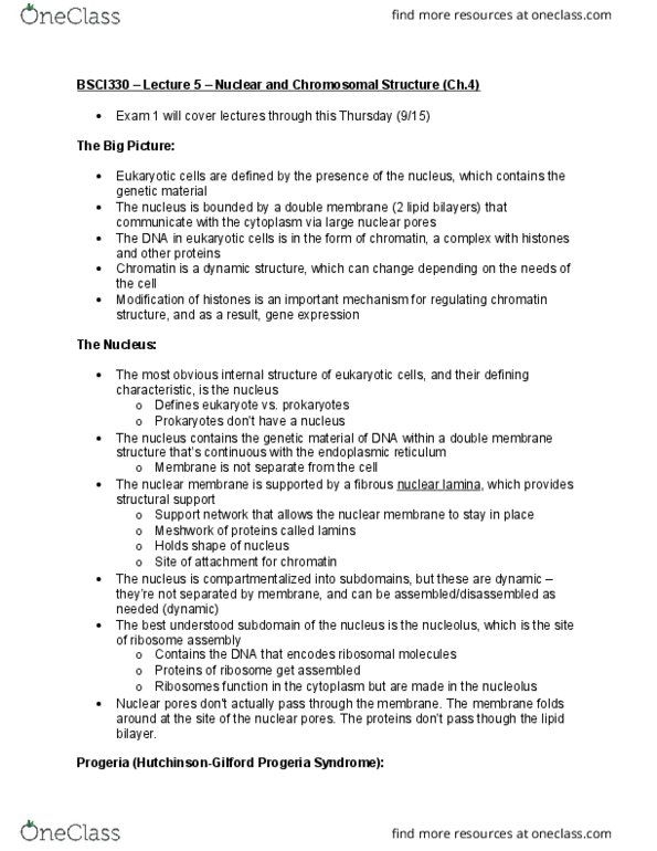
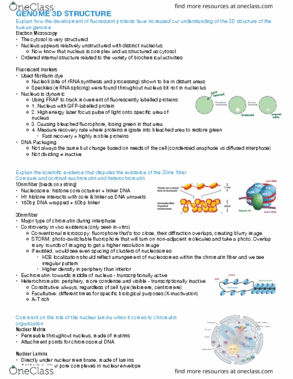
^that picture is from wednesday so i don"t know if we are responsible for that on the exam because the picture shows what the dna looks like when not wrapped around the histone. I think it is a picture of dna spilling out of a bacterium. If that is the case, we have seen the picture before and he did talk about it. Yeah but i"m pretty sure it was after lamina also. the 2nd question show the structure/indicate composition of the nuclear fibrous lamina. like what can we possibly draw other than the 2 proteins (lamin a and lamin b) We can draw the two bilayers and show that fibrous lamina is attached to the inner layer by bonding to intermembrane proteins. I think the answer is that you just put gold beads on the molecules and watch where they go/stay. Gold beads on trnas attached to molecules with nls. I guess just making an educated guess here.