BIO120H1 Lecture 20: lecture 20


www.notesolution.com

cell shape and function.
- Importantly, the nucleus is compartmentalized. Here, separating the
cytoplasm from the nucleus, there is a nuclear membrane shown in the
slide, the nuclear envelope.
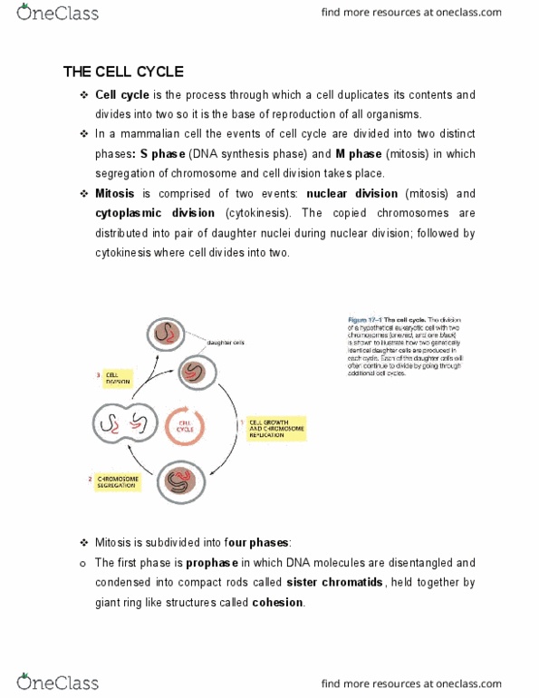
- Prophase is the first stage of mitosis and there are two main things that
start to happen, two main changes that occur.
- One is that sister chromatids start to condense, they align and they form
microtubule attachment sites. That is shown in the nucleus outlined in the
slide. Here in brown we see the DNA, these DNA double helices have been
replicated so now we have two copies of these so they were replicated
during interphase. Now we see these two pieces of DNA, each of them is
called a chromatid. The two chromatids are starting to connect together and
form one unit. Additionally, they form attachment sites for microtubules at
these red marks called kinetochore.
- At the same time this is happening, in the cytoplasm the centrosomes
begin to organize this bipolar microtubule spindle. Importantly, notice that
the microtubules and the DNA, they are still separated by a nuclear
envelope so they’re kept away from one another at this stage.
! Packaged and tagged to interact with microtubules
- Now to look at how these two steps happen.
- So we can think of this stage as preparing the DNA for mitosis and
preparing the microtubules for mitosis.
- For the DNA, there are basically 2 steps. One step is packaging them up
so instead of being very loose strands of DNA filling the nucleus, they are
packaged into solid packages that can be transported within the cell in an
effective way. So they’re packaged up and the other thing that is done to
them is that there is a tag put on them that is recognized by microtubules.
- There is basically 2 steps here, they’re packaged and tagged to interact
with microtubules.
! Individually condensed by condensins
! Glued together by complexes called cohesins
- This packaging occurs when all the DNA is duplicated in S phase, there
are 2 copies of each chromosomes and these remain associated as sister
chromatids.
- During prophase, each chromatid is individually condensed by condensins
and that is shown in this picture here, this is one DNA coil, one of these
sister chromatids. This DNA in interphase would be very loose and filling
the cell but here, these condensins are basically coiling this up and
packaging it together sort of like how you would coil up a hose in your
backyard. It connects them together making them a tighter package.
- In this picture right here, we are looking at 2 different sister chromatids.
The chromatids remain attached here called the centromere so that is an
important part of the chromosomes there, where those 2 chromatids remain
attached. We can see that they are each individually condensed by these
condensins & here in pink we can see the condensins running along each
sister chromatid. They are separately compacted by these condensins. These
condensed sister chromatids are glued together by complexes called
cohesions.
- This cartoon up here, these are the two sister chromatids. These cohesins
are connecting these condensed chromatids together into one large package.
If we fit this into this cartoon right here, the cohesins would be running
down the center attaching one sister chromatid to the other sister chromatid
so they’re all one large unit.
www.notesolution.com

! Microtubules
! Kinetochores
- So that is packing these chromosomes, now they’re also tagged to interact
with microtubules. This tag for the microtubule attachment, this forms at
the centromere region of the chromosomes. This is where the two sister
chromatids remain attached to one another and these sites are specifically
called kinetochore so that is shown here in this diagram so this is a protein
complex that binds to these chromosomes and can also bind to the
microtubules. So this tags them to interact with microtubules and these
microtubules as we’ll see later will drag these chromosomes apart.
- That is organizing the DNA in the nucleus and as that DNA is being
organized in the nucleus, the microtubules in the cytoplasm are being
organized to form the mitotic spindle.
- If we go outside into the cytoplasm, the mitotic spindle is being organized
by the centrosomes. So we’ve learned about the centrosomes before, here is
the centrosomes structure right here. Deep inside the centrosomes there are
2 centrioles & then surrounding those centrioles are hundreds of different
proteins. The key proteins on the surface of these centrosomes are the
gamma tubulin ring complexes. These gamma tubulin ring complexes act
as templates to nucleate microtubule growth so they’ll start to nucleate
microtubule growth & MTs will now shoot out from these centrosomes.
- This is an important place & another thing we learned about microtubules
is that at these plus ends of these microtubules here that are emanating out
from the centrosomes, we know that plus ends of microtubules display
dynamic instability so they shoot out, they retract back, they shoot out, they
retract back so now we have this center in the cell that is shooting out
microtubules in all directions starting to explore the cytoplasm.
- Then the chromosomes that are right now in the nucleus are forming tags
on them that will bind to those microtubules so we have chromosomes that
are tagged to interact with microtubules and now we have this robust search
and capture mechanism that is developing outside the nucleus.
! Microtubule asters
- A key thing in this diagram there is only one of these centrosomes present
and to form a spindle with two ends to it, to separate the chromosomes to
the two ends of the cell, these centrosomes need to be replicated. So the
centrosomes duplicate before M phase and each of these will nucleate
microtubule growth and this produces what are called microtubule asters.
- They are called asters because of the star-like configurations of the
microtubules emanating out in every direction. That is shown right here, the
cell would be born with one centrosome from its previous division, it then
has to replicate and then the two asters will move to opposite ends of the
cell during prophase so they’ll initially be right next to each other as they
replicate and then they migrate to opposite ends of the cell and then more
and more microtubule nucleation from these centrosomes will generate a
robust microtubule array that makes up this very large spindle and then they
have the potential to interact with the chromosomes inside.
www.notesolution.com


36
BIO120H1 Full Course Notes
Verified Note
36 documents
Document Summary
Here, separating the cytoplasm from the nucleus, there is a nuclear membrane shown in the slide, the nuclear envelope. Prophase is the first stage of mitosis and there are two main things that start to happen, two main changes that occur. One is that sister chromatids start to condense, they align and they form microtubule attachment sites. That is shown in the nucleus outlined in the slide. Here in brown we see the dna, these dna double helices have been replicated so now we have two copies of these so they were replicated during interphase. Now we see these two pieces of dna, each of them is called a chromatid. The two chromatids are starting to connect together and form one unit. Additionally, they form attachment sites for microtubules at these red marks called kinetochore. At the same time this is happening, in the cytoplasm the centrosomes begin to organize this bipolar microtubule spindle.