ANAT 261 Lecture : Lab 8. Oral Cavity.doc

15 Mar 2012
School
Department
Course
Professor

Document Summary
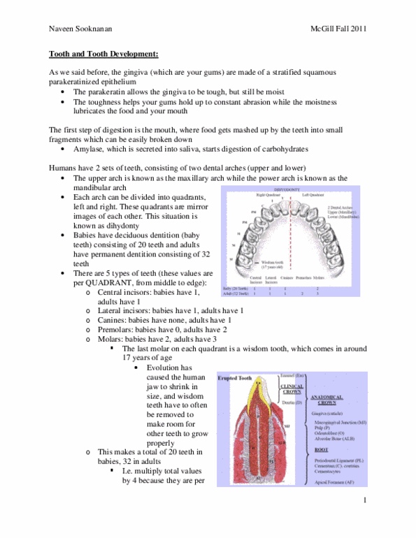
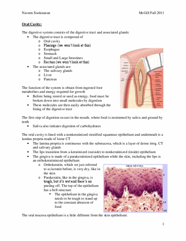
Erupted tooth: clinical crown: consists of enamel and dentin, won"t see enamel in lab! Above gingiva (gums: anatomical crown: clinical crown + gingiva continues until point where enamel layer ends. Gingiva = parakeratinized stratified squamous epithelium (makes gum tough and resistant: root and associated structures: *mucogingival junction parakeratin disappears and epithelium becomes non-keratinized oral mucosa (continues to. Root: pulp: loose ct, blood vessels, lymphatics, nerves, odontoblasts: line pulp & secrete dentin (as pre- dentin . stains pale pink) Columnar shape, nuclei at base: dentin: has lined appearance from cannaliculi (stains darker pink, cementum: runs down outside edge of dentin. [innermost to outermost: developing pulp, odontoblasts, predentin and dentin, enamel, enamel organ: forms enamel. Stratum intermedium: ameloblasts = tall columnar cells, 1-3 polygonal cell layers, star shaped ; lots of intercellular space. **ide and ode merge at the root sheath! Lab 8: oral cavity: 1-2 squamous cell layers. Epithelium covering the tongue = non-keratinized stratified squamous epithelium: filiform papilla: