OTHY102 Study Guide - Final Guide: Acromioclavicular Joint, White Matter, Endomysium


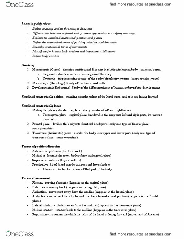
Anatomy: The study of the structure of living organisms.
• Further categorised into gross anatomy (study of large scale structures) and microscopic anatomy
(study of microscopic structures).
Regions of the body
Anatomical Position (Shown above)
• Body erect
• Feet apart
• Palms facing forward
• Thumbs pointing away from body
Planes of the Body
• Median Plane/Mid Sagittal Plane: Midline plane dividing the body exactly into left and right halves
• Sagittal Plane: Divides the body into left and right parts (not halves)
• Coronal/Frontal Plane: Splits the body into front and back
• Transverse or horizontal: Divides the body into top and bottom
find more resources at oneclass.com
find more resources at oneclass.com

Types of Movements
• Active movements = produced by active muscle
contraction.
Flexion: Bending a joint/decreasing the
angle between the bones of a joint.
Extension: Straightening a joint.
Abduction: Moving the joint away from
the midline of the body.
Adduction: Moving the joint towards the
midline of the body.
Rotation: Turning the bone around its axis medially (toward the body) or laterally (away
from the body)
Supination: External rotation of the radiohumeral joint. Wrist and palm face up.
Pronation: Internal rotation of the radiohumeral joint. Wrist and palm face down.
Inversion: Turning the sole of the foot inward
Eversion: Turning the sole of the foot outward
• Passive movements = Movement produced by an external force
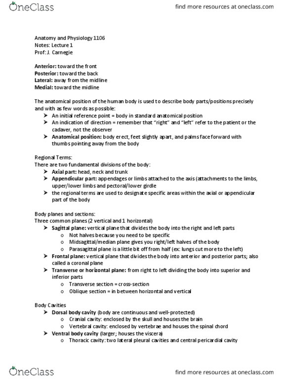
Directional Terms
• Superior: Closer to the top of the head
• Inferior: Closer to the feet
• Anterior: More in front
• Posterior/Dorsal: More in back
• Medial: Closer to the medial plane/midline of the body
• Lateral: Further from the medial plane/closer to the sides of the body
• Proximal: Limbs being closer to the medial plane or root of the limb than another structure in the
limb
• Distal: Limbs being farther from the medial plane/root of the limb than another structure in the
limb.
find more resources at oneclass.com
find more resources at oneclass.com

Bones
Divisions of the Skeletal System
• Axial; Bones of the vertebral column, thorax, pelvis & skull
• Appendicular skeleton; Bones of the limbs and limb girdles (attach limbs to body)
Functions of the Human Skeleton
• Provides shape and support
• Enables movement
• Protection for internal structures
• Production of blood cells
• Storage of minerals
Bone Tissue
• External: Compact bone
• Internal: Spongy bone, medullary cavity
Classifying Bones by Shape
•Short bone: eg. Tarsal
•Long bone: eg. Metatarsal.
Features of a long bone include the diaphysis and epiphysis.
Diaphysis: Long, cylindrical structure forming the main shaft. Thick
layer of compact bone over a thin layer of spongy bone. Hollow
central part of the bone forming the medullary/marrow cavity.
Covered in periosteum.
Epiphysis: Rounded parts located at the ends of a long bone. Filled
with spongy bone & red bone marrow. Covered in compact bone.
Joint surfaces covered in cartilage
• Sesamoid bone: eg. Patella. Function of a sesamoid bone is to increase
efficiency of the muscle. Not directly connected to any other bones.
• Flat bone: eg. Scapula
• Irregular bone: eg. Vertebra
Bone Markings
Features of a bone that function to:
• Strengthen
• Provide passages through the bone
• Promote bone to bone articulation
• Provide attachment sites for muscles
• Provide landmarks
find more resources at oneclass.com
find more resources at oneclass.com
Document Summary
Anatomy: the study of the structure of living organisms: further categorised into gross anatomy (study of large scale structures) and microscopic anatomy (study of microscopic structures). Anatomical position (shown above: body erect, feet apart, palms facing forward, thumbs pointing away from body. Types of movements: active movements = produced by active muscle contraction. Flexion: bending a joint/decreasing the angle between the bones of a joint. Abduction: moving the joint away from the midline of the body. Adduction: moving the joint towards the midline of the body. Rotation: turning the bone around its axis medially (toward the body) or laterally (away from the body) Inversion: turning the sole of the foot inward. Eversion: turning the sole of the foot outward: passive movements = movement produced by an external force. Divisions of the skeletal system: axial; bones of the vertebral column, thorax, pelvis & skull, appendicular skeleton; bones of the limbs and limb girdles (attach limbs to body)