OTHY200 Study Guide - Final Guide: Angiography, Midbrain, Degenerative Disease


OTHY200 Neuroscience and Neurodevelopment –
Stream B Revision
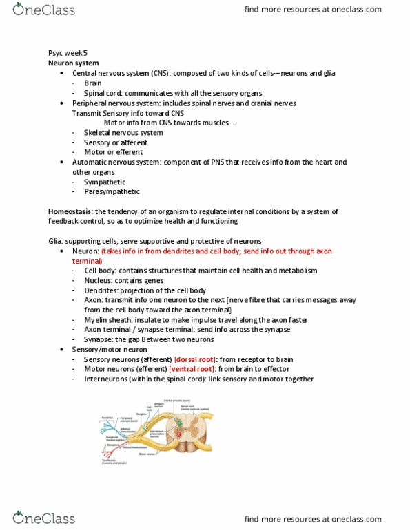
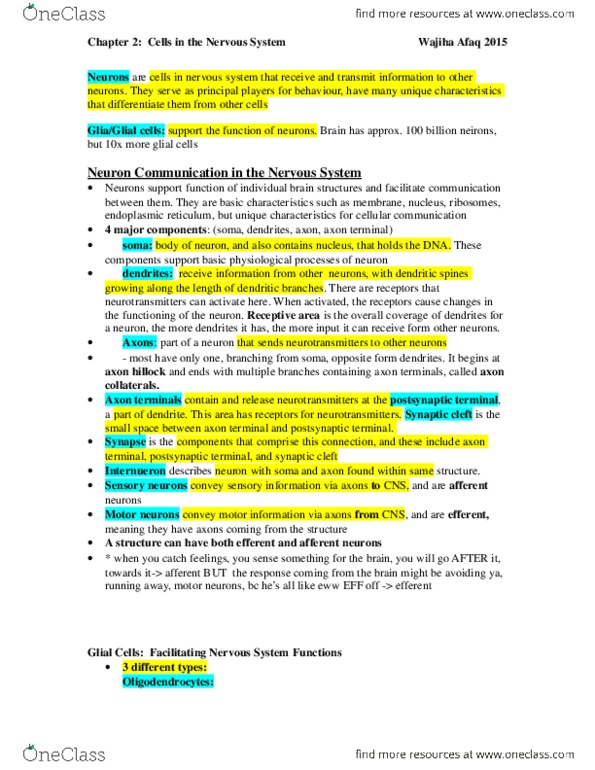
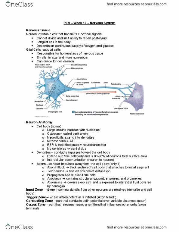
Neurons – Functional unit of the NS.
Cell body – Houses organelles of cell. Receptive part of the neuron (has dendrites)
Axon – Action potential travels down the axon towards synapse
Axon terminal – Neurotransmitters are released from AT at the synapse
Neurons are classified according to the direction in which they transmit impulses
Afferent = Sensory neuron. Towards CNS
Efferent = Motor neuron. Away from CNS
Directional Terms
Ventral = Anterior (Front) – Usually motor
Dorsal = Posterior (Back) – Usually sensory
Rostral = Superior (Up)
Caudal = Inferior (Down)
Cerebral Hemispheres
- Largest past of the CNS
find more resources at oneclass.com
find more resources at oneclass.com

OTHY200 Neuroscience and Neurodevelopment –
Stream B Revision
- Broken into left and right hemispheres
- Has gyri (hills) and sulci (valleys)
- Outer part is cerebrum is grey matter
- Deep part is white and grey matter
Grey and White Matter
• Grey matter:
- Cell bodies of neurons
• White matter:
- Axonal parts of neuron
- Connects spinal cord to the brain or connects parts of the brain
- 3 classifications of the fibres; projection, commissural and association
- Projection fibres = Sensory/motor fibres that connect the brain with the brainstem and spinal cord
- Commissural fibres = Connect the two hemispheres
- Association fibres = Connect areas within one hemisphere (Ipsilateral: Same sided)
Lobes of the Brain
There are four lobes in each cerebral hemisphere
• Frontal
- Motor functioning
- Broca’s area
• Parietal
- Somatosensory functioning
• Temporal
- Hearing
- Wernicke’s area
• Occipital
-Vision
Major Landmarks of the Brain
- Longitudinal fissure = Splits left and right hemisphere
- Central sulcus = Splits frontal and parietal lobe
- Lateral sulcus = Splits temporal lobe from lobes above
- Pre-central gyrus = Location of PMC
- Post-central gyrus = Location of PSC
- Corpus callosum = C shaped structure inside the brain, linking hemispheres
- Parieto-occipital sulcus = Divides parietal and occipital lobes
- Calcarine sulcus = Runs horizontally through occipital lobe
find more resources at oneclass.com
find more resources at oneclass.com

OTHY200 Neuroscience and Neurodevelopment –
Stream B Revision
Brodmann Areas
- Numerical system for locating the functional areas of the cortex
Brainstem
- Stalk of the brain connecting spinal cord to brain
- Houses cranial nerves numbered III - XII
- Contains pathways/tracts
- Regulates fundamental bodily functions; heartrate, swallowing, vomiting, coughing
- Divided into 3 parts: Midbrain, pons, medulla
• Midbrain:
- Uppermost part of the brainstem
- Houses CN nuclei III and IV (eye movement)
- Houses red nucleus and substantia nigra
• Pons:
- Passage for motor information from the cerebral cortex into the cerebellum via peduncles
- Houses CN nuclei V (face sensation and chewing) and VII (facial expression)
• Medulla:
- Lowermost part of the brainstem
- Houses CN nuclei X (parasympathetic, IX, X, XI (ambiguous) and XII (tongue movement)
- Decussation point for corticospinal tract and dorsal column
Disorders in the Brainstem
- A brainstem lesion may cause a mix of ipsilateral and contralateral (opposite sided) signs
- Cranial nerves supply the ipsilateral face and neck but
- Many vertical tracts decussate in the brainstem to supply the contralateral body
- Brainstem lesions may also interfere with vital functions and consciousness (pons and medulla)
find more resources at oneclass.com
find more resources at oneclass.com
Document Summary
Axon action potential travels down the axon towards synapse. Axon terminal neurotransmitters are released from at at the synapse. Neurons are classified according to the direction in which they transmit impulses. Outer part is cerebrum is grey matter. Deep part is white and grey matter. Cell bodies of neurons: white matter: Connects spinal cord to the brain or connects parts of the brain. 3 classifications of the fibres; projection, commissural and association. Projection fibres = sensory/motor fibres that connect the brain with the brainstem and spinal cord. Commissural fibres = connect the two hemispheres. Association fibres = connect areas within one hemisphere (ipsilateral: same sided) There are four lobes in each cerebral hemisphere: frontal. Longitudinal fissure = splits left and right hemisphere. Central sulcus = splits frontal and parietal lobe. Lateral sulcus = splits temporal lobe from lobes above. Corpus callosum = c shaped structure inside the brain, linking hemispheres. Parieto-occipital sulcus = divides parietal and occipital lobes.