PHYS1008 Final: mri instrumentation, coils

2 Aug 2018
School
Department
Course
Professor
Document Summary
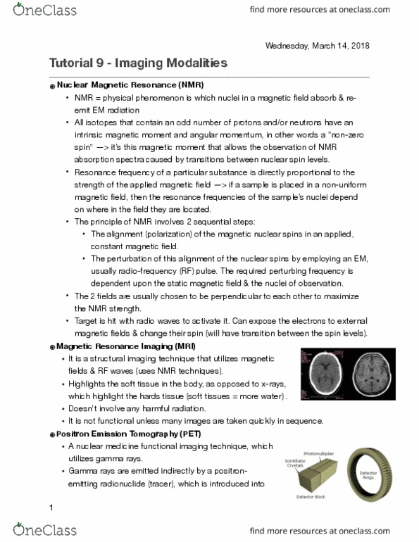
Imaging modality based on nuclear magnetic resonance: produces cross sectional images, visualises and analyses a variety of tissue characteristics, blood flow and metabolic functions. Interactions between static magnetic fields, radio waves and atomic nuclei: energy source = radio frequency, signal = intrinsic spin angular momentum of hydrogen gives you the signal, in a frequency domain, not spatial so it must be converted. Absorption of energy occurring when nuclei of atoms or molecules within an external magnetic field are exposed to radiofrequency energy (a radio wave) at a specific frequency called larmor of resonance frequency. Any nucleus with an odd number of protons or neutrons are able to produce a signal. However, mri is primarily applied to hydrogen because: its high sensitivity for its nmr signal, high abundance in the body (85% - in h20 molecules) The presence of change in the h nucleus (proton) leads to nuclear magnetism, the nucleus with charge will behave as a bar magnet does.