PSYC10003 Study Guide - Final Guide: Central Nervous System, Sodium Channel, Axon Terminal

16 Jun 2018
School
Department
Course
Professor

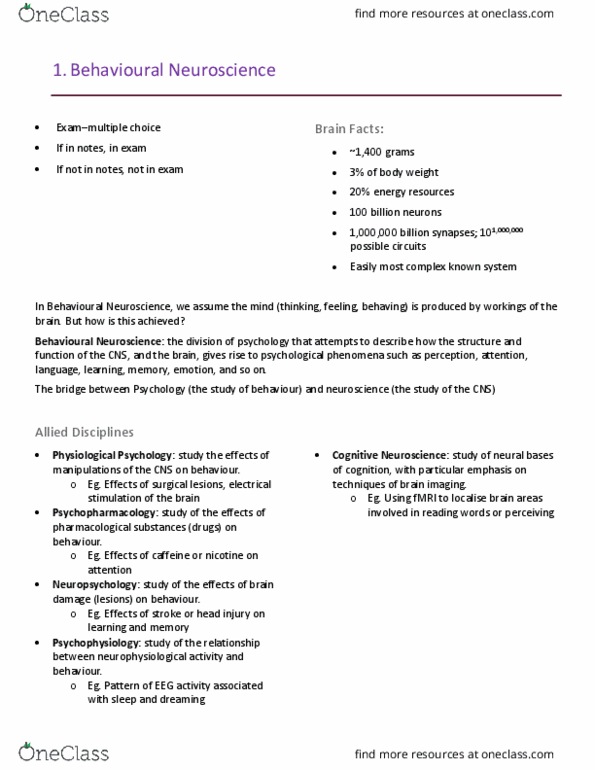
The human brain constitutes only 3% of our body weight, but uses 20% of our energy resources
It is made up of around 100 billion neurons
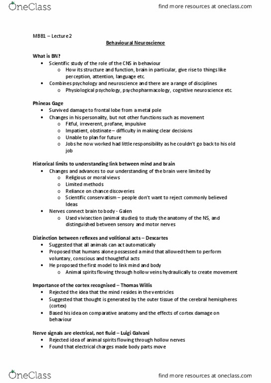
Behavioural Neuroscience – the division of psychology that attempts to describe how the
structure and function of the central nervous system (CNS), and the brain in particular, gives rise
to psychological phenomena such as perception, attention, language, learning, memory,
emotion, and so on.
Phineas Gage:
o Tamping iron through the skull, as result, Gage’s personality changed
Fitful, profane, impatient, obstinate, issues holding employment
Mind and souls was believed to be in heart in ancient cultures
Historical views of the brain based on:
o Religious beliefs
o Methods available to study it
o Chance discovery (serendipity)
o Strength under criticism
460BCE Hippocrates first to suggest brain as command center of body
130CE – Vivisection by Galen. Made connection between nerves and function.
1514: Andreas Vesalius. Revived Vivisection.
o Discovered two cerebral cortices
o Made good drawings of brain
o Showed the meninges, blood vessels, folding of brain
o The folded outer layer of brain tissue (cortex), composed of gyri (convolutions) and sulci
(grooves)
1596: Descartes proposes relaxes, suggests only humans have ability to react voluntarily.
o viewed the mind and body as distinct entities (i.e., he was a dualist)
o first to suggest that there was a link between the physical brain and the non-physical
mind.
o Thought animal spirits moved through ventricles to body.
1621: Thomas Willis, rejected idea that mind resides in ventricles.
o Suggests thought stems from cortex.

1737: Galvani rejects idea of animal spirits passing through hollow nerves.
o Found that electric charge made frogs leg muscle contract
o Suggests that nerves must be coated in fat to prevent electricity from leaking out.
1758: Franz Joseph Gall suggested several distinct organs of thought in brain that indicated
personality. Phrenology. Cortical localization of function.
1861: First Solid evidence of brain modularity. Paul Broca’s patient unable to speak after
damage to left frontal lobe
1870: Precise locations of brain function. Eg. To stimulate specific muscles.
20th century: Egas Monis introduced prefrontal leucotomy for relief of psychiatric disorders.
Consequences of disconnecting frontal lobes showed to be inconsistent, often severe.
Methods of study driven our knowledge:
Surgical and accidental lesions
Animal studies
Modularity: the mind is the product of the brain.
The neuron is the basic information-processing and information receiving unit of the nervous system.
Neurons form complex networks within the nervous system, but they are not directly connected with
one another; they are separated by a tiny gaps called synapses, across which chemicals called
neurotransmitters are passed. Neurons come in many different shapes and sizes. Almost all have four
basic structures or regions:
1) Cell body– contains the nucleus (genetic material) and internal organelles necessary for cell
maintenance.
2) Dendrites – the tree-like branches that allow neurons to communicate with one another. Dendrites
receive information from other neurons.
3) Axon – a long, slender fiber that carries signals from the cell body. The signal carried by an axon is an
action potential, which as we shall see later is a wave of electrical potential that begins at the cell body
and travels down the axon to the terminal buttons.
4) Terminal buttons) – small knobs at the ends of the many branches of axons. These structures play a
critical role in transmitting information from one neuron to another, by secreting neurotransmitters
which pass across the synaptic gap and can either excite or inhibit the next neuron in the chain.
Neurons may receive information from the terminal buttons of many other neurons, and may
themselves send information to many other neurons via their own terminal buttons. Terminal buttons
may form synapses with the cell body or the dendrites of other neurons.

Neurons constitute only half the volume of the CNS; the other half is made up of various other cells,
collectively known as glial cells (or glia), that play an important role in providing physical support for
neurons and in supplying them with oxygen and nutrients. There are several types of glial cell:
Astrocytes provide physical support for neurons and also do housekeeping jobs (cleaning up waste,
providing nutrients to neurons, maintaining the correct chemical composition of the extracellular fluid
that surrounds neurons). Some astrocytes literally crawl around the CNS, cleaning away the debris from
dead neurons, a process called phagocytosis. After removal of the dead neurons, other astrocytes will
take their place, thus maintaining a supportive structure for nearby cells. Oligodendrocytes also provide
physical support to neurons, but most importantly they provide the insulating myelin sheath that
surrounds the axon. This prevents unwanted cross-talk between neighboring axons. Most, but not all,
axons are myelinated. Microglia are the smallest glial cells. They act as phagocytes, like some astrocytes;
they also act as the brain’s immune system, attacking invading micro-organisms. Microglia are largely
responsible for inflammation after brain damage. Schwann cells perform the same function as
oligodendrocytes, only they do this in the peripheral nervous system (PNS). They create a myelin sheath
around the axons of neurons in the PNS. Oligodendrocytes: form a tube of myelin around the axon of
neurons in the CNS. This tube is made up of a series of segments of myelin, each roughly 1 mm long and
with a small gap of uncoated axon between them. These gaps are called Nodes of Ranvier (after their
discoverer) Schwann cells: form a tube of myelin around the axons of neurons in the PNS. The tube is
made of segments, and each segment consists of one entire Schwann cell. Schwann cells also possess a
special function not shared by oligodendrocytes: when there is damage to an axon, they digest the
remaining portion of the fibre and then align themselves into a hollow cylinder, to act as a guide for any
axonal stump that resprouts after damage. This process helps reconnect axons with the muscles and
sense organs with which they were originally connected
What is the nature of the signal transmitted by a neuron?
Basically it is an electrical process which involves movement across the axon membrane of electrically
charged molecules called ions.
resting membrane potential, is about -70 millivolts
Depolarization - A very rapid reversal of the membrane potential of an axon is called an action
potential. The action potential constitutes the basic message that is transmitted down an axon from the
cell body to the terminal buttons
There are four ions that are crucial to the resting membrane potential, two with a positive
charge (cations) and two with a negative charge (anions). These are:
o sodium (Na+);
o chloride (Cl- );
o potassium (K+)
o organic anions (which are proteins, A-)