BIOM2020 Study Guide - Final Guide: Urethral Sphincters, Abdominal Wall, Prostatic Urethra

Document Summary
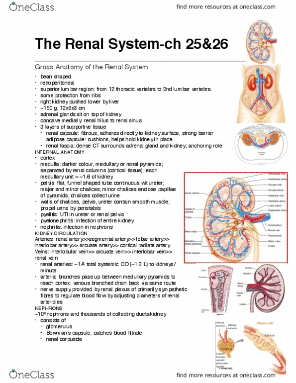
Left renal artery longer than the right due to descending aortic trunk in the way. Right kidney (starts at lumbar v) lower than the left (starts at thoracic v to ~l3) due to the position of the liver. Held in position by adipose tissue (no ligamentous attachments) Structure and function: suprarenal /adrenal glands sit superior to kidney. Regulates blood"s inorganic ion balance (na, k+, po42-) Balances acid-base by changing the rates of h+ and ammonium secretion. Renal fascia: dense irregular tissue -> anchors kidney to surroundings. Adipose capsule: fat + adipose -> cushioning and insulation. Renal capsule: dense connective tissue -> maintains shape and protects from pathogens. Sequence for urine elimination: collecting duct -> papillary duct -> minor calyx -> major calyx -> renal pelvis -> ureter -> urinary bladder. Minor and major calices located in renal sinus; as well as renal pelvis, renal fat and blood vessels. Hilum: contains renal vein, artery and ureter (ant. to post.