ANHB1102 Study Guide - Final Guide: Cardiac Output, Capillary, Action Potential

3 Jun 2018
School
Course
Professor

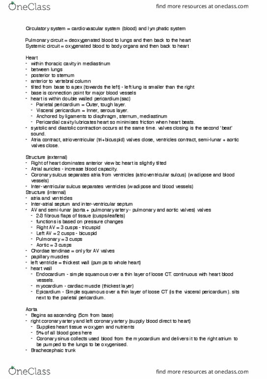
TOPIC THREE: THE CARDIOVASCULAR SYSTEM
Location of the Heart:
The heart is located in the thorax, found in the region between the lungs, called
the mediastinum. Since the heart is in the central position it is said to be in the
middle mediastinum.
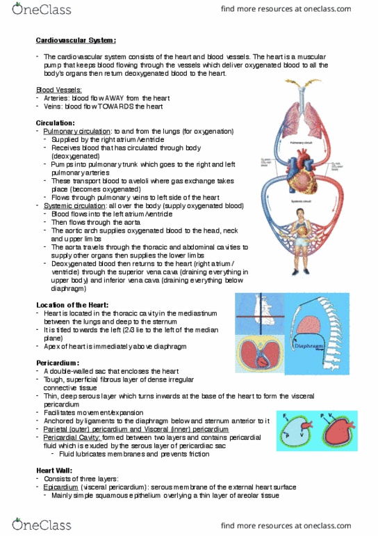
Pericardium:
Surrounding the heart is a substance called the
pericardium. There are two types of pericardium –
fibrous pericardium (structure F) and serous
pericardium. The serous pericardium is like a sac
wrapped around the heart, lined by epithelium and secreting watery fluid to
facilitate movement. There are two layers to the serous pericardium – the
parietal layer (structure P) and the visceral layer (structure V).
External Features of the Heart:
Internal Features of the Heart:
find more resources at oneclass.com
find more resources at oneclass.com

Development of the Heart:
Development of the heart begins during embryogenesis. Mesodermal masses
form the blood and blood vessels while cardiogenic mesoderm at the cranial end
of the embryo will form the structures of the heart. Cranial folding moves the
heart to the thoracic position and the heart tube is formed. The tail end of the
heart tube forms veins while the cranial end will form the arteries. At this stage,
the atrium is posterior while the ventricles are anterior. The heart must supply
blood even when it develops, so it begins beating by the end of week three.
From weeks four to eight, and beyond, there is partitioning of the heart, the
development of right and left sides. The lungs are not yet functional, so blood
will flow from a passage in the partition from the right atrium to left atrium until
birth. At birth, the passage in partition closes and systemic and pulmonary
circulation is separated.
Blood Vessels:
While there are a number of blood vessels carrying out varying tasks, all blood
vessels have a common basic plan. They contain a smooth inner surface of
endothelium. They contain elastic connective tissue so flow and pressure can
find more resources at oneclass.com
find more resources at oneclass.com
Document Summary
The heart is located in the thorax, found in the region between the lungs, called the mediastinum. Since the heart is in the central position it is said to be in the middle mediastinum. Surrounding the heart is a substance called the wrapped around the heart, lined by epithelium and secreting watery fluid to pericardium. There are two types of pericardium facilitate movement. There are two layers to the serous pericardium the fibrous pericardium (structure f) and serous pericardium. The serous pericardium is like a sac parietal layer (structure p) and the visceral layer (structure v). Mesodermal masses form the blood and blood vessels while cardiogenic mesoderm at the cranial end of the embryo will form the structures of the heart. Cranial folding moves the heart to the thoracic position and the heart tube is formed. The tail end of the heart tube forms veins while the cranial end will form the arteries.