PHYL2001 Study Guide - Final Guide: Hemoglobin, Carbamino, Vital Capacity


Respiratory Physiology
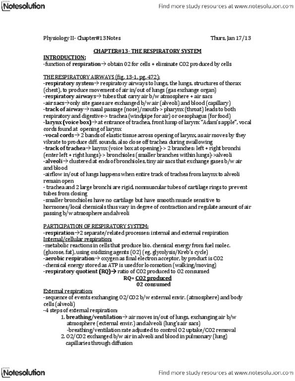
Anatomy of the Respiratory System:
• Respiratory Airways
o Tubes that carry air between the atmosphere and alveoli
o
o
o
o
o
o
o
o
o
o Airway
must always be open
to allow airflow
o Trachea and larger bronchi
▪ Fairly rigid
▪ Nonmuscular tubes
▪ Encircled by cartilaginous rings to prevent compression
o Smaller bronchioles
▪ No cartilage
▪ Smooth muscle
▪ Innervated by autonomic nervous system to regulate air
flow
• Alveoli
o Clusters of thin walled, inflatable sacs at the terminating
bronchioles
o Walls → single layer of flattened Type 1 cells
o Surrounded by a network of pulmonary capillaries → also a single
wall thick
o Thin barrier between alveolus and capillary wall facilitates gas
exchange
o Alveoliar air-pulmonary blood interface presents an increased
surface area for exchange
o Lungs contain 500 million alveoli
o 5% alveolar suface → type 2 alveolar cells → secrete surfactant
▪ Surfactant → facilitates lung expansion
o Pores of John → exist in walls between adjacent alveoli → allows
airflow between adjoining alveoli
• Lungs
o 2 lungs → divided by lobes supplied by one of the bronchi
find more resources at oneclass.com
find more resources at oneclass.com

o Lung tissue consists of
▪ Series of highly branched airways
▪ Pulmonary blood vessels
▪ Large quantities of elastic connective tissue
o Only smooth muscle
o Changes in lung volume are brought about by changes in the
dimensions of the thoracic cavity
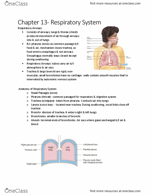
• Thoracic cavity
o Outer chest wall → ribs, sternum, thoracic vertebrae
o Rib cage → protection for heart and lungs
o Diaphragm → floor of thoracic cavity → separates thoracic from
abdominal cavity
o Pleural sac → separates lungs from thoracic wall
• Pleurae
o Visceral pleura → surrounds lungs
o Parietal pleura → lines chest
o Pleural space → contains fluid
o Function of pleural fluid
▪ Reduction of friction
▪ Creation of a pressure gradient
▪ Compartmentalization
Function of the Respiratory System:
• Conducting zone
o Warm and humidify air
o Distribute gas
• Respiratory zone
o Site of gas exchange
• Non-respiratory functions
o Route for water loss and heat elimination
o Enhances venous return
o Maintain normal acid-base balance
o Enables speech, singing and other vocalizations
o Defends against inhaled foreign matter
Respiratory Pressures:
• Boyles Law →
(at constant temperature)
• Charles Law → (at constant pressure)
• Gas Law →
=
• All measured in cm H2O → 0.74 mmHg (or 1 mmHg = 1.36 cm H2O)
• Atmospheric pressure
o Pressure exerted by the weight of the air in the atmosphere on
objects on the Earths surface
o Sea level → 760 mmHg
o Decreases with increasing altitude
• Transairway pressure (Pta)
find more resources at oneclass.com
find more resources at oneclass.com

o Prevents airway collapsing during forced expiration
o Pta = Paw - Ppi
• Transpulmonary pressure (Pl)
o Prevents lungs collapsing
o Pl = Pa - Ppl
• Intra-alveolar pressure
o Pressure within alveoli
o For air to flow into the lungs during inspiration, atmospheric
pressure > intra-alveolar pressure
o For air to flow out of the lungs during expiration, atmospheric
pressure < intra-alveolar pressure
• Intra-pleural pressure
o Pressure in pleural sac
o Allows lungs to stay open
o Slightly less than atmospheric pressure → less than 760mmHg
o Reaches approximately 754mmHg during inspiration
Respiratory Cycle:
• Inspiration
o Onset of inspiration → contraction of inspiratory muscles
▪ Diaphragm
▪ External intercostal muscles
o Before inspiration → muscles are relaxed
o Contraction of inspiratory muscles → increase size of thoracic
cavity
o Contraction of diaphragm causes it to move downward →
innervated by the phrenic nerve
o Contraction of external intercostals enlarges thoracic cavity in
lateral and anteroposterior dimensions → innervated by
intercostal nerves
o Before inspiration, intra-alveolar pressure = atmospheric pressure
o As thoracic cavity expands, pressure drops → air flows into lungs
o Deeper inspiration achieved by contracting diaphragm and
external intercostals more forefully and including accessory
inspiratory muscles
▪ Sternocleidomastoid
▪ Sclaneus
▪ Both raise sternum and first 2 ribs → larger flow of air into
lungs → deeper breath
• Expiration
o Onset of inspiration → relaxation of inspiratory muscles
o Chest wall and stretched lungs recoil to normal size because of
elastic properties
o Intra-alveolar pressure increases → air leaves lungs
o Ceases when intra-alveolar pressure = atmospheric pressure
o Quiet breathing → expiration is passive
o Forced expiration → contraction of active expiratory muscles
▪ Internal intercostal muscles
find more resources at oneclass.com
find more resources at oneclass.com
Document Summary
Intra-alveolar pressure: pressure within alveoli, for air to flow into the lungs during inspiration, atmospheric pressure > intra-alveolar pressure, for air to flow out of the lungs during expiration, atmospheric pressure < intra-alveolar pressure. Intra-pleural pressure: pressure in pleural sac, allows lungs to stay open, slightly less than atmospheric pressure less than 760mmhg, reaches approximately 754mmhg during inspiration. Internal intercostal muscles: pulls ribs downward and inward, flattens chest wall, abdominal muscles most important. Increase in intra-abdominal pressure exerts an upward force on the diaphragm: pushes it to thoracic cavity, lung size reduced, greater differential between intra-alveolar and atmospheric pressure more air leaves. Spirometry: tidal volume, volume of air entering or leaving lungs in a single breath, ~ 500ml. Inspiratory reserve volume: extra volume of air that can be maximally inspired over and above typical resting tidal volume, achieved by maximal contraction of diaphragm, external intercostal muscles and accessory inspiratory muscles, ~ 3000ml.