PHYL2002 Study Guide - Final Guide: Exosphere, Exocytosis, Lidocaine


Fast Signaling in the Nervous System
Introduction to the Nervous System:
• Central nervous system → brain and spinal cord
• Peripheral nervous system → nerves and ganglia
o Visceral afferents → blood pressure, pain, osmolarity
o Somatic afferents → touch/temperature, proprioception, balance
o Special senses → vision, hearing, taste, smell
o Autonomic nervous system → sympathetic/parasympathetic
nerves
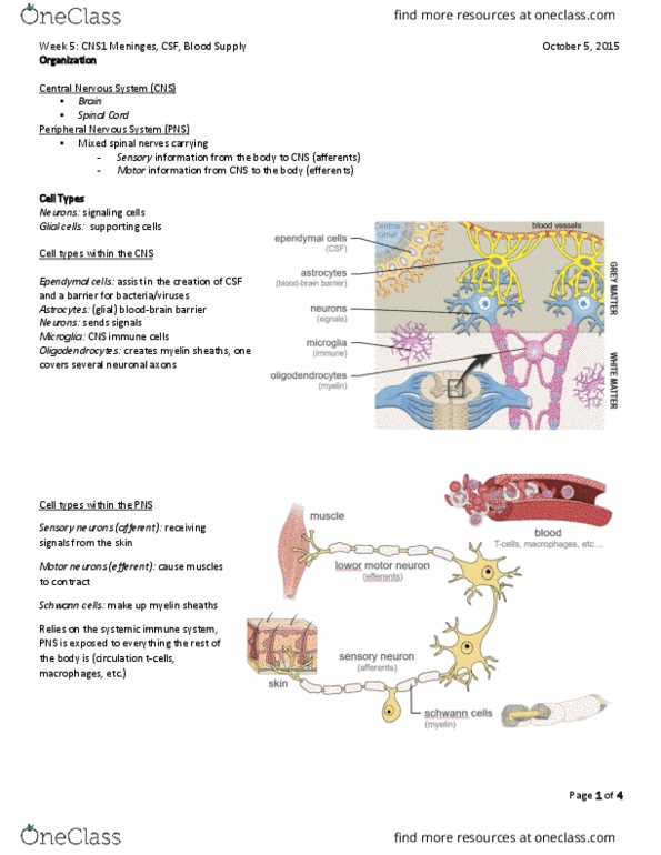
• Glial cells
o Support cells
o Most abundant
o Microglia
▪ Phagocytes
▪ Arise from macrophages outside nervous system
▪ Embryologically unrelated to other nervous system cells
o Macroglia
▪ Oligodendrocytes → formation of myelin sheaths in CNS
▪ Schwann cells → formation of myelin sheaths in PNS
▪ Astrocytes → blood-brain barrier, reuptake transmitters
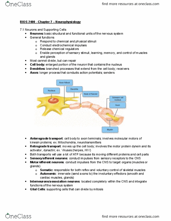
o Myelin → lipid-rich, impermeable to ions
o Nodes of Ranvier → not covered by myelin
• Neurons
o Main signaling cells
o Function
▪ Structural support and insulation of neurons
▪ Myelin sheaths → oligodendrocytes and Schwann cells
▪ Scavenging dead cells → microglia
▪ Uptake of released neurotransmitters, buffer for excess K+
▪ Radial glia direct migration of developing neurons
▪ Blood-brain barrier → astrocytes and endothelial cells
▪ Trophic support for neurons
o Classification
▪ Number of neurites
▪ Size
▪ Shape
▪ Neurochemistry
▪ Location
▪ Connectivity
o Structure
▪ Cell body → soma
▪ Projections from the soma → neuritis
▪ Dendrites → neuritis which receive input
▪ Axons → transmit signals long distances
▪ Terminals → where neurotransmitters are released from
find more resources at oneclass.com
find more resources at oneclass.com

▪ Neurotransmitters → chemicals that signal between nerve
cells
o Nerve signaling
▪ Nerve to nerve connection → synapses
▪ Nerve to gland/muscle → junction
▪ Pre-synaptic cell sends the signal
▪ Post-synaptic cell receives the signal
• Neuronal signaling
o Types of synapses
▪ Axosomatic synapses
▪ Axodendritic synapses
▪ Axoaxonic synapses
o Resting membrane potential → -60 mV
o Due to
▪ Unequal distribution of electrically charged ions
▪ Selective permeability of membrane of these ions
o In nerve cells membrane potential can be quickly altered by
changes in permeability to certain ions
o Intracellular recordings enable measurement of membrane
potential
o Changes in potential are used to transmit information within the
nervous system
o Graded potentials signal over short distances
o Action potentials signal over long distances
o Vm decays due to leakage of electrically charged ions
o Distance at which Vm has decayed to 37% of original value at
current injection defines length constant
o Length constant is a measure of efficiency of passive speed
• Axon hillock
o Threshold for action potential lowest at Axon Hillock
o Axon hillock highest density of sodium channels
o Effectiveness of synaptic connection depends on the distance
length constant
o Integration of all excitatory and inhibitory inputs
o The less a passive signal decays, the more likely to generate an
action potential
Ion Channels:
• Allow lipophobic ions to travel through lipid membrane
• Ion channels are selective for specific ions
• Most ion channels are gated
• Movement of ions is passive
• Structure
o Primary structure → sequence of amino acids
o Secondary structure → alpha helix
o Tertiary structure → 3D folding of polypeptide
o Quaternary structure → different polypeptides bound together
find more resources at oneclass.com
find more resources at oneclass.com

o Membrane spanning a-helices contain hydrophobic amino acid
residues
o Connected by loops of hydrophilic residues
o 4 domains containing 6x membrane spanning hydrophobic a-
helices
o Aqueous pores formed by one of the a-helices
o Another a-helix (S4) contains voltage sensor → center of channel
• Diversity is created due to availability of many different subunits
o Heterooligomers → distinct subunits
o Homooligomers → single subunits
• Ion channels are selective due to
o Size of pore
▪ Small throat will not let large ions through
o Electrical charge of chains of amino acids that enter pore
▪ Negative charged throat repels anions makes channel
cation selection
• In solutions ions have hydrogen bonds to water → hydrated shell
• Most hydrogen bonds to water replaced by bonds to channel amino acids
• Gating
o Opening → conformational change occurs in one region of channel
o Inactivation → blocking particle swings into and out of channel
mouth
o Ligand gated → required binding of a chemical
o Voltage gated → requires a voltage change across the membrane
o Mechanically gated → requires stretching or some displacement
• Ligand gated channels
o Direct
▪ Channel opens in response to binding of the ligand to
receptor
▪ Energy from ligand binding drives channel gating towards
an open state
▪ May be neurotransmitters or hormones
▪ Bind on extracellular side of channel
o Antagonist can inhibit binding of endogenous ligand
▪ Curare → blocks nicotinic Ach receptor
▪ Lidocaine → local anaesthetic, binds to domain IV of Na+
channel, inhibiting action potential generation
▪ Tetrodotoxin → found in puffer fish, binds to voltage
generated Na+ channels preventing action potential
generation
o Indirect
▪ Channel opens in response to a second messenger signal
activated by a neurotransmitter
▪ Second messenger acts on intracellular side of channel →
couples receptor to the ion channel
o Nicotinic receptors in muscle
▪ Found on skeletal muscle
▪ Opened by Ach from nerve
▪ 5 subunits for the channel pore
find more resources at oneclass.com
find more resources at oneclass.com