PSYC340 Study Guide - Final Guide: Calcitriol, Monoglyceride, Myenteric Plexus

24 Jun 2018
School
Department
Course
Professor

1. Describe the location, anatomy, histology and functions of the small intestine.
A. Small intestine - Most digestion and absorption of nutrients occur in a long tube
called the small intestine. Because of this, its structure is specially adapted for these
functions. Its length alone provides a large surface area for digestion and absorption,
and that area is
further increased by circular folds, villi, and microvilli
o1. Segmentations mix chyme with digestive juices and bring food into
contact with mucosa for absorption; peristalsis propels chyme through
small intestine.
o2. Completes digestion of carbohydrates, proteins, and lipids; begins and
completes digestion of nucleic acids.
o3. Absorbs about 90% of nutrient and water that pass through digestive
system.
A.I. Anatomy
A.II. Duodenum
A.III. The first part of the small intestine is the duodenum, the shortest region,
and is retroperitoneal. It starts at the pyloric sphincter of the stomach
and is in the form of a C-shaped tube that extends about 25 cm (10 in.)
until it merges with the jejunum. Duodenum means “12”; it is so named
because it is about as long as the width of 12 fingers
A.IV. Jejunum
A.V. The jejunum is the next portion and is about 1 m (3 ft) long and extends
to the ileum. Jejunum means “empty,” which is how it is found at death.
A.VI. Ileum/Ileocecal sphincter
A.VII. The final and longest region of the small intestine, the ileum, measures
about 2 m (6 ft) and joins the large intestine at a smooth muscle
sphincter called the ileocecal sphincter (valve)
A.VIII. Histology
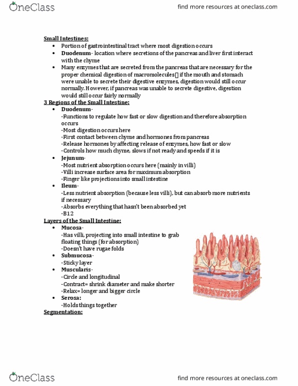
A.VIII.a. Mucosa - The mucosa is composed of a layer of
epithelium, lamina propria, and muscularis mucosae.
A.VIII.b. Absorptive cells - Absorptive cells of the epithelium release
enzymes that digest food and contain microvilli that absorb nutrients
in small intestinal chyme.
A.VIII.c. Goblet cells - Also present in the epithelium are goblet
cells, which secrete mucus
A.VIII.d. Intestinal glands or crypts of Lieberkuhn - Cells lining the
crevices form the intestinal glands, or crypts of Lieberkühn, and
secrete intestinal juice (to be discussed shortly). Besides absorptive
cells and goblet cells, the intestinal glands also contain paneth cells
and enteroendocrine cells.
A.VIII.e. Paneth cells - Paneth cells secrete lysozyme, a bactericidal
enzyme, and are capable of phagocytosis. Paneth cells may have a
role in regulating the microbial population in the small intestine.
find more resources at oneclass.com
find more resources at oneclass.com

A.VIII.f. Secretin /Cholecystokinin (CCK)/ Glucose dependent
insulinotropic peptide (GIP) – types of enteroendocrine cells
A.VIII.g. Solitary lymphatic nodules - Solitary lymphatic nodules are
most numerous in the distal part of the ileum
A.VIII.h. Aggregated lymphatic follicles or Peyer’s patches - Groups
of lymphatic nodules referred to as aggregated lymphatic follicles, or
Peyer’s patches, are also present in the ileum. The muscularis
mucosae of the small intestinal mucosa consists of smooth muscle
A.VIII.i. Submucosa / Duodenal glands or Brunner’s glands
A.VIII.j. The submucosa of the duodenum contains duodenal
glands, also called Brunner’s glands, which secrete an alkaline
mucus that helps neutralize gastric acid in the chyme.
A.VIII.k. Muscularis - Sometimes the lymphatic tissue of the lamina
propria extends through the muscularis mucosae into the submucosa.
The muscularis of the small intestine consists of two layers of smooth
muscle
A.VIII.l. Serosa - The outer, thinner layer contains longitudinal
fibers; the inner, thicker layer contains circular fibers. Except for a
major portion of the duodenum, which is retroperitoneal, the serosa
(or visceral peritoneum) completely surrounds the small intestine
A.VIII.m. Circular folds - Circular folds or plicae circulares are folds
of the mucosa and submucosa
A.VIII.m.i. These permanent ridges, which are about 10 mm (0.4 in.)
long,
begin near the proximal portion of the duodenum and end
at about the midportion of the ileum. Some extend all the
way around the circumference of the intestine; others
extend only part of the way around. Circular folds enhance
absorption by increasing surface area and causing the
chyme to spiral, rather than move in a straight line, as it
passes through the small intestine
A.VIII.n. Villus (plural is villi) - Also present in the small intestine are
villi (! tufts of hair), which are fingerlike projections of the mucosa
that are 0.5–1 mm long
A.VIII.o. The large number of villi (20 – 40 per square mm) vastly
increases the surface area of the epithelium available for
absorption and digestion and gives the intestinal mucosa a velvety
appearance.
A.VIII.p. Lacteal
A.VIII.q. Each villus (singular form) is covered by epithelium and has
a core of lamina propria; embedded in the connective tissue of
the lamina propria are an arteriole, a venule, a blood capillary
network, and a lacteal, which is a lymphatic capillary
find more resources at oneclass.com
find more resources at oneclass.com

A.VIII.r. Nutrients absorbed by the epithelial cells covering the
villus pass through the wall of a capillary or a lacteal to enter
blood or lymph, respectively.
A.VIII.s. Microvillus (plural is microvilli)
A.VIII.t. Besides circular folds and villi, the small intestine also has
microvilli, which are projections of the apical (free) membrane of
the absorptive cells. Each microvillus is a 1-"m-long cylindrical,
membrane-covered projection that contains a bundle of 20–30
actin filaments
A.VIII.u. Because the microvilli greatly increase the surface area of
the plasma membrane, larger amounts of digested nutrients can
diffuse into absorptive cells in a given period. The brush border
also contains several brush-border enzymes that have digestive
functions
A.VIII.v. Brush border
A.VIII.w. When viewed through a light microscope, the microvilli
are too small to be seen individually; instead they form a fuzzy
line, called the brush border, extending into the lumen of the
small intestine
A.VIII.x. Intestinal juice
A.VIII.y. About 1–2 liters (1–2 qt) of intestinal juice, a clear yellow
fluid, is secreted each day. Intestinal juice contains water and
mucus and is slightly alkaline (pH 7.6). The alkaline pH of intestinal
juice is due to its high concentration of bicarbonate ions (HCO3 -).
Together, pancreatic and intestinal juices provide a liquid medium
that aids the absorption of substances from chyme in the small
intestine
A.VIII.z. Brush border enzymes: dextrinase, maltase, sucrase,
lactase, peptidases (including aminopeptidase and dipeptidase),
nucleosidases an phosphatases
A.IX. Mechanical digestion - The two types of movements of the small
intestine—segmentations and a type of peristalsis called migrating motility
complexes—are governed mainly by the myenteric plexus
A.X. Segmentations
A.XI. Segmentations are localized, mixing contractions that occur in portions of
intestine distended by a large volume of chyme.
A.XII. Segmentations mix chime with the digestive juices and bring the particles
of food into contact with the mucosa for absorption; they do not push
the intestinal content along the tract. A segmentation starts with the
contractions of circular muscle fibers in a portion of the small intestine,
an action that constricts the intestine into segments.
A.XIII. Next, muscle fibers that encircle the middle of each segment also
contract, dividing each segment again.
find more resources at oneclass.com
find more resources at oneclass.com
Document Summary
Describe the location, anatomy, histology and functions of the small intestine, small intestine - most digestion and absorption of nutrients occur in a long tube called the small intestine. Because of this, its structure is specially adapted for these functions. Its length alone provides a large surface area for digestion and absorption, and that area is further increased by circular folds, villi, and microvilli: 1. Segmentations mix chyme with digestive juices and bring food into contact with mucosa for absorption; peristalsis propels chyme through small intestine: 2. Completes digestion of carbohydrates, proteins, and lipids; begins and completes digestion of nucleic acids: 3. Absorbs about 90% of nutrient and water that pass through digestive. The first part of the small intestine is the duodenum, the shortest region, and is retroperitoneal. It starts at the pyloric sphincter of the stomach and is in the form of a c-shaped tube that extends about 25 cm (10 in. ) until it merges with the jejunum.