PHGY 209 Study Guide - Final Guide: Inferior Vena Cava, Heart Valve, Pulmonary Artery

15 Feb 2018
School
Department
Course
Professor
Document Summary
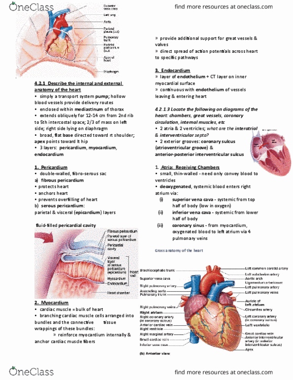
Heart: enclosed in double sac called pericardium containing serous fluid (friction-free, walls composed of 3 layers: superficial epicardium, myocardium (cardiac. Muscle and connective tissue), endocardium (endothelium, squamous epithelium: 4 chambers: 2 superior atria, 2 inferior ventricles, atrium: receiving blood, small thin walled. Right atrium receives oxygen-poor blood (deoxygenated) from above heart via superior vena cava, from below the heart via inferior vena cava and blood from the myocardium via coronary sinus. Left atrium receives oxygen- rich (oxygenated) blood from both lungs via 4 pulmonary veins: ventricles: discharging blood from heart to arteries, large thick walled especially the left ventricle. Inside: internal ridges and cone-like papillary muscle attached to atrio-aventricular valves. Right ventricle sends oxygen-poor blood to the lungs (pulmonary circulation) via pulmonary trunk leading into two pulmonary arteries. Left ventricle sends oxygen-rich blood via aorta to systemic circulation and to the myocardium via right and left coronary arteries: heart valves: one-way flow, close due to change in pressure.