PHGY 209 Study Guide - Final Guide: Neural Tube, Incus, External Carotid Artery

18 Jun 2016
School
Department
Course
Professor
Document Summary
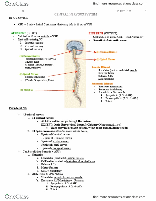
Sulci: fissures: central sulcus: separate frontal lobe from somatosensory. White matter: myelanited axons connecting various portion of brain, connect basal nuclei (ganglia), which are cells bodies involve in motor control. Cranial nerves: 12 in total, 10 enters the brain, 2 do not go through => olfactory and optic nerve. When cutting part of thoracic nerve: loose somatosensation and motor control in thin strip of upper abdominal wall, shoulder and chest. Dorsal horn: at back receives sensory inputs. Ventral horn: in front contains cell body from motor neurons: dorsal root ganglion. Where cell bodies for sensory afferent (accumulate in enlargement near the vertebrae) Week 1: blastocyst with inner cell mass. Week 3: blastocyst with embryonic disk: neural plate. Week 4: 3 vesicles (forebrain, midbrain, hindbrain) are developing and the rest elongates itself. Fully developed: forebrain: cerebral hemispheres and thalamus, midbrain: midbrain, hindbrain: pons, medulla, cerebellum.