PSYC 2410 Midterm: PSYC-2410-Lectures-5-8-Review

27 Jun 2018
School
Department
Course
Professor

PSYC 2410 Midterm 2 – Lecture Materials
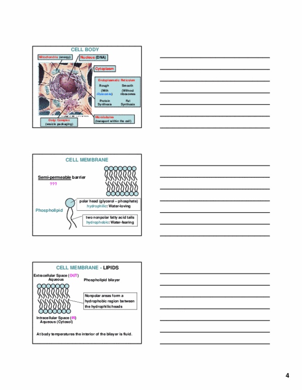
Lecture 5: Excitable Cell Membranes
- The cell membrane has a semi-permeable barrier
o Not everything can enter through it. Ions/molecules can enter through it either by being
small enough to fit, or by using membrane-imbedded channels/proteins to help enter
the cell
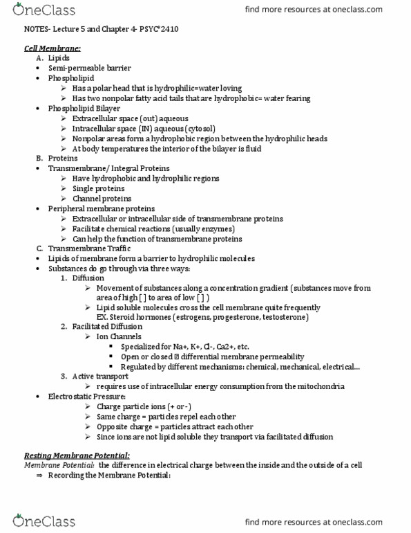
o Structure: Phospholipid
o These tails form the PHOSPHOLIPID BILAYER. The heads face outwards in both directions
which allows aqueous interactions while the tails remain in the middle and form a
hydrophobic region
o At body temperatures, the interior of the bilayer is fluid
- Membrane Proteins:
o “transmembrane” or “integral” proteins
Both hydrophobic and hydrophilic regions
Includes SIGNAL proteins and CHANNEL proteins
o “peripheral” membrane proteins (i.e. non-transmembrane)
Extracellular or intracellular side of transmembrane proteins
Act as facilitators. Things they facilitate are:
Chemical reactions (usually act as enzymes)
Functions of transmembrane proteins
- The hydrophobic part of the cell membrane can make it difficult for a water-soluble molecule to
pass through, but different methods exist for this to be able to occur.
o Active Transport (requires energy consumption via mitochondria)
o Passive processes: no energy consumed
- Diffusion: movement of substance along a concentration gradient
o Moving from an area of high concentrationlow concentration is passive transport
(uses no energy) while low conchigh conc is active transport.
o Can be done with lipid soluble molecules (e.g., steroid hormones)
- Ions are charged particles
o Concentration gradient and electrostatic pressure act together to determine the
distribution of ions…
o **IONS ARE NOT LIPID SOLUBLE**
o Therefore, ion channels exist on the cell membrane
Form of “facilitated diffusion”
Are specialized for different ions, e.g., sodium (Na), chlorine (Cl), calcium (Ca),
etc
These channels can be open or closed so that the membrane’s permeability can
be changed (regulated by chemical, mechanical, electrical mechanisms)
- Charges near the cell membrane
o
o Electrical properties of the cell membrane are determined by the uneven distribution of
charges (i.e. Ions) on either side
o Sodium (Na+) and Chlorine (Cl-) are more concentrated OUTSIDE THE CELL
o Potassium (K+) and protein ions (-) are more concentrated INSIDE THE CELL
o Overall, the INSIDE=MORE NEGATIVE than outside
o This differential concentration can be maintained because the cell membrane can
decide how easily it can let in ions…
Impermeable to electrically charged proteins (-)
Not very permeable to sodium ions (Na+)
Relatively permeable to potassium ions (K+)
Very permeable to chloride ions (Cl-)
- Axon in NaCl (i.e. salt) solution
o Membrane potential: difference in electrical charge between the inside and the outside
of a cell (related to ion distribution)
o Neuronal Resting potential = -70mV (milliVolts)
i.e. a cell at -70 mV is POLARIZED
- Hodgkin and Huxley in 1950 calculated the electrostatic charge needed to keep the various ions
in the location where, at rest, they are in greater concentration:
o Cl-: 70 mV 70-70 = 0 (subtract 70 because resting potential is -70)
o K+: 90 mV 90-70 = 20 mV
o Na+: 50 mV 50 + 70 = 120 mV
We are adding the 70 in sodium’s case because both the electrostatic pressure
and concentration gradient are acting in the same direction
o Hodgkin and Huxley also showed that:
K+ ions “leak out” and Na+ ions go into the cell, even at rest although the
distribution still stays relatively stable because there are pumps to take care of
this
- SODIUM-POTASSIUM (Na-K) PUMP:
o Active transport mechanism
o Uses energy from mitochondria (ATP)
o 3 Na:2 K
3 sodium ions pumped out; 2 potassium ions pumped in
electrostatic charge tells it to stay outside
because it is more attracted to the negative
external environment

- Synapses on dendritic spines or the cell body
o Synaptic neurotransmitters can cause changes in the potential of post-synaptic neurons.
May depolarize the receptive membrane (e.g. -70 -67 mV)
May hyperpolarize the receptive membrane (e.g. -70 -72 mV)
- Depolarization Excitatory Postsynaptic Potentials
- Hyperpolarization Inhibitory Postsynaptic Potentials
o Both types of polarization are small changes that can increase or decrease the chance of
the cell firing an action potential
- Post Synaptic Potentials:
o Graded responses: this means that the extent to which the potential is
de/hyperpolarized depends on the intensity of the signal that caused them.
o The transmission of postsynaptic potentials is passive and occurs extremely quick
(pretty much instantaneous)
o The transmission of postsynaptic potentials is decremental. This means that as the
potentials travel through the neuron, they decrease in amplitude
- Post Synaptic Potentials ADD TOGETHER (aka integration)
o Integration: adding or combining a number of individual signals into one overall signal
o Spatial Summation: at the same time in different locations (these potentials can cancel
each other out if they are equal magnitude in opposite direction)
o Temporal Summation: the integration of neural signals that occur at different times at
the same synapse
- Action potential:
o Summation threshold of excitation (approx.. -65 mV) neuron fires (action potential
triggered)
o “all or none”… if you reach -66 mV, the threshold hasn’t been reached so nothing will
happen; and no matter how far above the threshold you go, it will fire at the same
intensity no matter what because the action potential has been reached (e.g., if the
Document Summary
The cell membrane has a semi-permeable barrier: not everything can enter through it. Ions/molecules can enter through it either by being small enough to fit, or by using membrane-imbedded channels/proteins to help enter the cell: structure: phospholipid, these tails form the phospholipid bilayer. The heads face outwards in both directions which allows aqueous interactions while the tails remain in the middle and form a hydrophobic region. The hydrophobic part of the cell membrane can make it difficult for a water-soluble molecule to pass through, but different methods exist for this to be able to occur. Membrane proteins: at body temperatures, the interior of the bilayer is fluid, transmembrane or integral proteins. Includes signal proteins and channel proteins: peripheral membrane proteins (i. e. non-transmembrane) Extracellular or intracellular side of transmembrane proteins. Are specialized for different ions, e. g. , sodium (na), chlorine (cl), calcium (ca), etc.